. 2020 Sep 1;28(4):385-398.
doi: 10.3727/096504020X15861709922491. Epub 2020 Apr 6.

Cylindromatosis Is Required for Survival of a Subset of Melanoma Cells

Affiliations

Cylindromatosis Is Required for Survival of a Subset of Melanoma Cells

Ting La et al. Oncol Res. .

Abstract

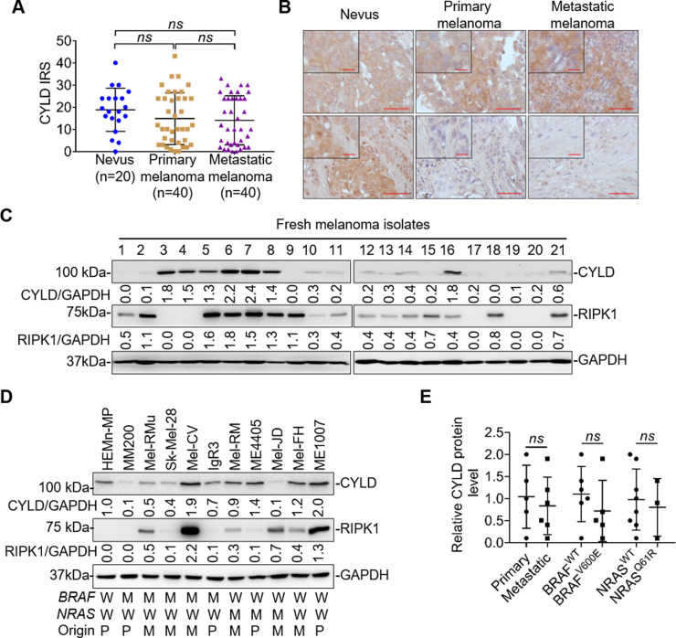
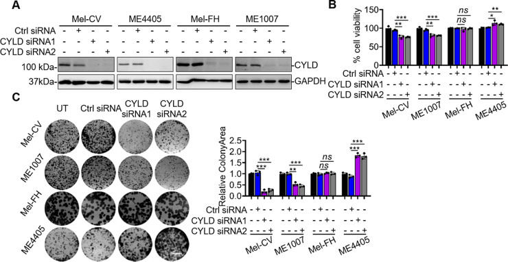
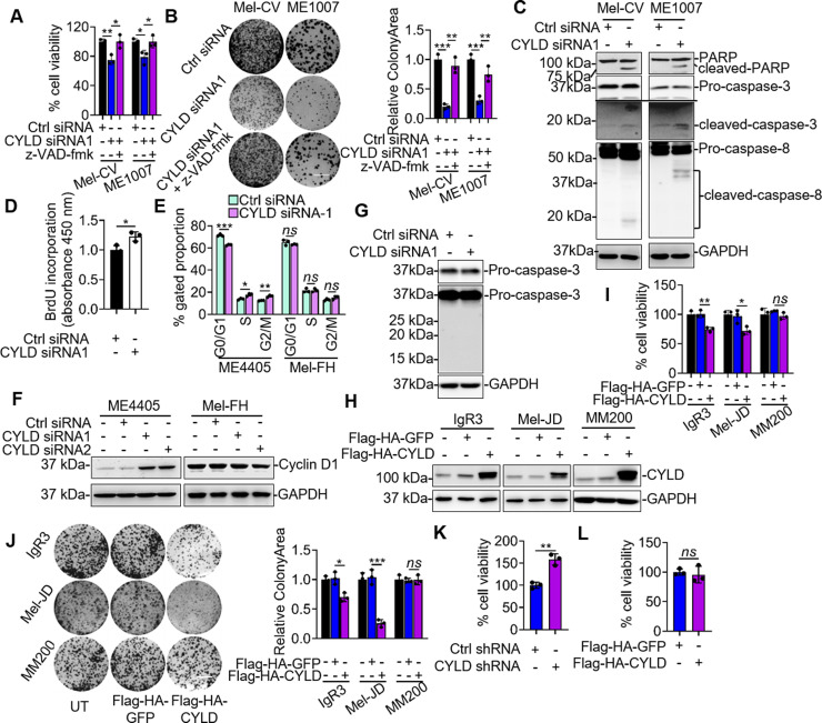
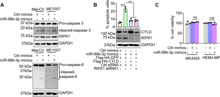
The deubiquitinase cylindromatosis (CYLD) functions as a tumor suppressor inhibiting cell proliferation in many cancer types including melanoma. Here we present evidence that a proportion of melanoma cells are nonetheless addicted to CYLD for survival. The expression levels of CYLD varied widely in melanoma cell lines and melanomas in vivo, with a subset of melanoma cell lines and melanomas displaying even higher levels of CYLD than melanocyte lines and nevi, respectively. Strikingly, although short hairpin RNA (shRNA) knockdown of CYLD promoted, as anticipated, cell proliferation in some melanoma cell lines, it reduced cell viability in a fraction of melanoma cell lines with relatively high levels of CYLD expression and did not impinge on survival and proliferation in a third type of melanoma cell lines. The decrease in cell viability caused by CYLD knockdown was due to induction of apoptosis, as it was associated with activation of the caspase cascade and was abolished by treatment with a general caspase inhibitor. Mechanistic investigations demonstrated that induction of apoptosis by CYLD knockdown was caused by upregulation of receptor-interacting protein kinase 1 (RIPK1) that was associated with elevated K63-linked polyubiquitination of the protein, indicating that CYLD is critical for controlling RIPK1 expression in these cells. Of note, microRNA (miR) profiling showed that miR-99b-3p that was predicted to target the 3-untranslated region (3-UTR) of the CYLD mRNA was reduced in melanoma cell lines with high levels of CYLD compared with melanocyte lines. Further functional studies confirmed that the reduction in miR-99b-3p expression was responsible for the increased expression of CYLD in a highly cell line-specific manner. Taken together, these results reveal an unexpected role of CYLD in promoting survival of a subset of melanoma cells and uncover the heterogeneity of CYLD expression and its biological significance in melanoma.


Conflict of interest statement

The authors declare no conflicts of interest.

Figures

Figure 1
Figure 1
Cylindromatosis (CYLD) is heterogeneously expressed in human melanocytic cells. (A) Quantification of CYLD expression in melanocytic tumors. Data shown are mean immunoreactive score (IRS) ± standard error of the mean (SEM). ns, p > 0.05, Kruskal–Wallis test. (B) Representative microphotographs of immunohistochemistry (IHC) staining of CYLD on melanocytic tissue sections. Scale bar: 100 μm. (C, D) Whole-cell lysates from melanocytes and melanoma cells were subjected to Western blotting. Data shown are representative of three individual experiments. W, wild-type; M, BRAFV600E or NRASQ61R mutation; P, primary melanoma; M, metastatic melanoma. (E) The relative abundance of CYLD was compared between primary (n = 5) and metastatic (n = 6), BRAFWT (n = 6) and BRAFV600E (n = 5), and NRASWT (n = 8) and NRASQ61R (n = 3) melanoma cell lines. Data were adapted from (D). ns, p > 0.05, Student’s t-test.
Figure 2
Figure 2
CYLD differentially regulates melanoma cell survival and proliferation. (A, B) Mel-CV, ME4405, Mel-FH, and ME1007 cells transfected with indicated CYLD short hairpin RNAs (siRNAs) were subjected to Western blotting (A) and CellTiter-Glo assays (B). Data shown are representative of three individual experiments (A) or means ± SEM (B). *p < 0.05; **p < 0.01; ***p < 0.001, Student’s t-test. (C) Mel-CV, ME1007, Mel-FH, and ME4405 cells transfected with indicated CYLD siRNAs were subject to clonogenic assays (left). Scale bar: 1 cm. Relative colony area were measured by ImageJ (right). Colony area in control cells was arbitrarily designated as 1. Data shown are representative of three individual experiments (left) or means ± SEM (right). n = 3. **p < 0.01; ***p < 0.001, Student’s t-test.
Figure 3
Figure 3
CYLD differentially regulates melanoma cell survival and proliferation in a cell line-dependent manner. (A, B) Mel-CV and ME1007 cell lines transfected with indicated siRNAs and with or without z-VAD-fmk were subject to CellTiter-Glo assays (A) and clonogenic assay (B). Data shown are representative of three individual experiments or means ± SEM. n = 3. *p < 0.05; **p < 0.01; ***p < 0.001, Student’s t-test. (C) Whole-cell lysates from Mel-CV and ME1007 transfected with indicated siRNAs were subjected to Western blotting. Data shown are representative of three individual experiments. (D) ME4405 cells transfected with indicated siRNA were subjected to 5-bromo-2′-deoxyuridine (BrdU) incorporation assays. Data shown are representative of three individual experiments or means ± SEM. n = 3. *p < 0.05. (E, F) ME4405 and Mel-FH cells transfected with indicated siRNAs were subjected to cell cycle distribution (E) and Western blotting (F). Data shown are representative of three individual experiments or means ± SEM. n = 3. *p < 0.05; **p < 0.01; ***p < 0.001; ns, p > 0.05, Student’s t-test. (G) Mel-FH cells were transfected with indicated siRNA and subjected to Western blotting. Data shown are representative of three individual experiments. (H–J) IgR3, Mel-JD, and MM200 cell lines transfected with indicated plasmids were subjected to Western blotting (H), CellTiter-Glo assays (I), and clonogenic assay (J). Scale bar: 1 cm. Data shown are representative of three individual experiments or means ± SEM. n = 3. *p < 0.05; **p < 0.01; ***p < 0.001; ns, p > 0.05, Student’s t-test. (K, L) HEMn-MP melanocytes transduced with indicated shRNAs (K) or cDNA (L) were subjected to CellTiter-Glo assays. Data shown are means ± SEM. n = 3. **p < 0.01; ns, p > 0.05, Student’s t-test.
Figure 4
Figure 4
CYLD is critical for protection against the receptor-interacting protein kinase 1 (RIPK1)-induced apoptosis in a subset of melanoma cells. (A) Mel-CV, ME1007, Mel-FH, and ME4405 cell lines transfected with indicated siRNAs were subjected to Western blotting. Data shown are representative of three individual experiments. (B) Immunoprecipitates with RIPK1 antibody from Mel-CV and ME1007 cells transfected with indicated siRNAs and plasmids were subjected to Western blotting. Data shown are representative of three individual experiments. (C) Whole-cell lysates from Mel-CV and ME1007 cell lines transfected with indicated plasmids with or without treatment with MG132 were subjected to Western blotting. Data shown are representative of three individual experiments. (D) Mel-CV and ME1007 cells transfected with indicated siRNAs were subjected to propidium iodide (PI)/annexin V staining assays. Data shown are representative of three individual experiments (left) or means ± SEM (right). n = 3. ***p < 0.001, Student’s t-test. (E) ME4405 and Mel-FH cells transfected with indicated siRNAs were subjected to CellTiter-Glo assays. Data shown are means ± SEM. *p < 0.05; **p < 0.01; ns, p > 0.05, Student’s t-test. (F) Whole-cell lysates from Mel-CV and Mel-FH cells were subjected to Western blotting. Data shown are representative of three individual experiments. (G) Mel-CV and ME1007 cells with CYLD knocked down by siRNA were transfected with the indicated plasmids were subjected to PI/annexin V staining assays (top) and Western blotting (bottom). Data shown are means ±SEM (top) or representative of three individual experiments (bottom). n = 3. **p < 0.01; ***p < 0.001, Student’s t-test.
Figure 5
Figure 5
Cell line-dependent regulation of CYLD by microRNA-99b-3p (miR-99b-3p). (A) Total RNAs from melanocytes and melanoma cells were subjected to quantitative polymerase chain reaction (qPCR) analysis. Data shown are means ± SEM. (B) Mel-CV, ME4405, Mel-FH, and HEMn-MP cells were treated with or without cycloheximide (CHX; 5 μg/ml) for indicated periods. Whole-cell lysates were subjected to Western blotting (left). Quantification of CYLD relative to glyceraldehyde-3-phosphate dehydrogenase (GAPDH) is also shown (right). Mean ± SEM, n = 3, Student’s t-test. (C) Top 10 miRNAs that expressed the lowest level in melanoma cell lines Mel-CV and ME1007 in comparison with melanocytes. (D) Total RNAs from melanocytes and melanoma cells were subjected to qPCR analysis. Data shown are means ± SEM. (E) A schematic illustration of base pairing between miR-99b-3p and the 3′-untranslated region (3′-UTR) of CYLD. (F) A schematic illustration of psiCHECK2-based luciferase reporter constructs. (G) Luciferase reporter activity measured in Mel-CV and ME1007 cells after cotransfection with indicated reporter constructs and miRNA mimics. Data shown are means ± SEM, n = 3. *p < 0.05; ***p < 0.001, Student’s t-test. (H) Luciferase reporter activity measured in Mel-CV and ME1007 cells after cotransfection with indicated reporter constructs and anti-miRNA oligonucleotides. Data shown are means ± SEM. n = 3. **p < 0.01; ***p < 0.001, Student’s t-test. (I) Luciferase reporter activity measured in ME4405 and Mel-FH cells transfected with the indicated reporter constructs with or without cotransfection of miR-99b-3p mimics or anti-miR-99b-3p oligonucleotides. Data shown are means ± SEM. n = 3. p > 0.05, Student’s t-test. (J, K) Whole-cell lysates from Mel-CV and ME1007 cells transfected with indicated miRNA mimics (J) or anti-miRNA oligonucleotides (K) were subjected to qPCR analysis (top) and Western blotting (bottom). Data shown are representative of means ± SEM or three individual experiments. **p < 0.01; ***p < 0.001; ****p < 0.00001, Student’s t-test. (L) Whole-cell lysates from ME4405 and Mel-FH cells transfected with indicated miRNA mimics (top) or anti-miRNA oligonucleotides (bottom) were subjected to Western Blotting. Data shown are representative of three individual experiments.
Figure 6
Figure 6
RIPK1 rescues apoptosis induced by miR-99b-3p in Mel-CV and ME1007 cells. (A) Whole-cell lysates from Mel-CV and ME1007 cells transfected with indicated miRNA mimics were subjected to Western blotting. Data shown are representative of three individual experiments. (B) Mel-CV cells were transfected with indicated miRNA mimics, and plasmids were subjected to Western blotting (bottom) and PI/annexin V staining assays (top). Data shown are representative of three individual experiments (bottom) or means ± SEM (top). n = 3. **p < 0.01; ***p < 0.001, Student’s t-test. (C) ME4405 and HEMn-MP cells transfected with indicated miRNA mimics were subjected to CellTiter-Glo assays. Data shown are means ± SEM. n = 3. Student’s t-test.

Similar articles

Cited by

References

    1. Luke JJ, Flaherty KT, Ribas A, Long GV. Targeted agents and immunotherapies: Optimizing outcomes in melanoma. Nat Rev Clin Oncol. 2017;14(8):463–82. - PubMed
    1. Gide TN, Quek C, Menzies AM, Tasker AT, Shang P, Holst J, Madore J, Lim SY, Velickovic R, Wongchenko M, Yan Y, Lo S, Carlino MS, Guminski A, Saw RPM, Pang A, McGuire HM, Palendira U, Thompson JF, Rizos H, Silva IPD, Batten M, Scolyer RA, Long GV, Wilmott JS. Distinct immune cell populations define response to anti-PD-1 monotherapy and anti-PD-1/anti-CTLA-4 combined therapy. Cancer Cell 2019;35(2):238–55.e6. - PubMed
    1. Franke V, Berger DMS, Klop WMC, van der Hiel B, van de Wiel BA, Ter Meulen S, Wouters M, van Houdt WJ, van Akkooi ACJ. High response rates for T-VEC in early metastatic melanoma (stage IIIB/C-IVM1a). Int J Cancer 2019;145(4):974–8. - PubMed
    1. Menzies AM, Long GV. Recent developments in melanoma therapy. JAMA Oncol. 2016;2(10):1259–60. - PubMed
    1. Kozar I, Margue C, Rothengatter S, Haan C, Kreis S. Many ways to resistance: How melanoma cells evade targeted therapies. Biochim Biophys Acta Rev Cancer 2019;1871(2):313–22. - PubMed

MeSH terms