. 2020 May 22;295(21):7529-7543.
doi: 10.1074/jbc.RA120.012617. Epub 2020 Apr 6.

Mutations in penicillin-binding protein 2 from cephalosporin-resistant Neisseria gonorrhoeae hinder ceftriaxone acylation by restricting protein dynamics

Affiliations

Mutations in penicillin-binding protein 2 from cephalosporin-resistant Neisseria gonorrhoeae hinder ceftriaxone acylation by restricting protein dynamics

Avinash Singh et al. J Biol Chem. .

Abstract

The global incidence of the sexually transmitted disease gonorrhea is expected to rise due to the spread of Neisseria gonorrhoeae strains with decreased susceptibility to extended-spectrum cephalosporins (ESCs). ESC resistance is conferred by mosaic variants of penicillin-binding protein 2 (PBP2) that have diminished capacity to form acylated adducts with cephalosporins. To elucidate the molecular mechanisms of ESC resistance, we conducted a biochemical and high-resolution structural analysis of PBP2 variants derived from the decreased-susceptibility N. gonorrhoeae strain 35/02 and ESC-resistant strain H041. Our data reveal that mutations both lower affinity of PBP2 for ceftriaxone and restrict conformational changes that normally accompany acylation. Specifically, we observe that a G545S substitution hinders rotation of the β3 strand necessary to form the oxyanion hole for acylation and also traps ceftriaxone in a noncanonical configuration. In addition, F504L and N512Y substitutions appear to prevent bending of the β3-β4 loop that is required to contact the R1 group of ceftriaxone in the active site. Other mutations also appear to act by reducing flexibility in the protein. Overall, our findings reveal that restriction of protein dynamics in PBP2 underpins the ESC resistance of N. gonorrhoeae.

Keywords: Neisseria gonorrhoeae; X-ray crystallography; antibiotic resistance; ceftriaxone; conformational change; extended-spectrum cephalosporins; oxyanion hole; penicillin-binding protein; peptidoglycan; protein dynamic.


Conflict of interest statement

The authors declare that they have no conflicts of interest with the contents of this article

Figures
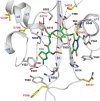
Figure 1.
Figure 1.
Mutations in PBP2 identified as contributing to ESC resistance of N. gonorrhoeae mapped onto the crystal structure of tPBP2WT acylated by ceftriaxone (30). Only the active-site region of PBP2 is shown, in which the protein is in ribbon form and colored gray, and sites of mutation are shown in yellow and labeled in red. Ceftriaxone (CRO) is shown with green bonds, and potential hydrogen bonds made by the antibiotic are indicated by dashed lines.
Figure 2.
Figure 2.
Mutations conferring ESC resistance affect both affinity and the rate of deacylation of tPBP2. A, isothermal titration calorimetry data for the interaction between ceftriaxone and S310A mutants of tPBP2. Upper panels show the binding isotherms for the wildtype (WT), 35/02, and H041 variants of tPBP2, and the lower panel shows the fit of the data for tPBP2WT by nonlinear regression with a single-site isotherm using NanoAnalyze software (TA Instruments). Three technical replicates were performed under identical conditions using 100 μm protein and 1000 μm ceftriaxone. B, deacylation rates (k3) for tPBP2WT- and tPBP2H041–acylated complexes. WT and H041 tPBP2 constructs were incubated with Bocillin-FL for an hour, after which ceftriaxone was added to a concentration of 3 mm (t = 0). Aliquots were removed at time intervals and separated by SDS-PAGE, and the amount of Bocillin-FL remaining bound was detected by UV illumination and quantified by densitometric scanning. The data were normalized for protein levels using Coomassie staining. Data were fit to first-order kinetics to derive the Bocillin-FL deacylation constant, k3. Experiments were performed in triplicate using protein samples from the same purification. For both experiments, the errors reported are the standard error of the mean.
Figure 3.
Figure 3.
Structures of tPBP235/02 and tPBP2H041 are highly similar. Shown is an overlay of the two backbones where tPBP235/02 is orange, and tPBP2H041 is green. The secondary structure nomenclature is the same as in Powell et al. (22).
Figure 4.
Figure 4.
Comparison of tPBP2WT and tPBP2H041 structures. A, backbones of the two structures in ribbon form are shown superimposed, where tPBP2WT is gray, and tPBP2H041 is green. Note differences in the β3–β4 and β2c–β2d loops. B, overlay of the active sites showing alteration of the Ser-362 side-chain rotamer in tPBP2H041. Distances in Ångstroms are indicated.
Figure 5.
Figure 5.
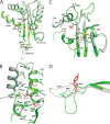
Impact of ESC resistance mutations on the structure of tPBP2H041. A, superimposition of tPBP2WT (gray) and tPBP2H041 (green) structures showing the positions of the seven critical mutations present in the active-site region of PBP2. Side chains of mutated residues are shown for both structures, and their Cα positions are indicated by red spheres for the tPBP2H041 structure. B–D show close-up views of the mutations. B, three mutations in helix α2. C, G545S on the β5–α11 loop and the T483S mutation on the α10–β3 loop. Note Ser-545 forms a potential hydrogen bond with Thr-498 in the structure of tPBP2H041 as indicated by a dashed line. D, F504L and N512Y mutations in the β3–β4 loop. For all panels, relevant side chains are shown in stick form and are colored according to structure. Mutations associated with ESC resistance are colored red and labeled in the same color. Mutations present in PBP2 from H041 but not directly implicated in ESC resistance are underlined.
Figure 6.
Figure 6.
Structure of tPBP2H041 acylated by the extended-spectrum cephalosporin ceftriaxone (CRO). Shown is an unbiased |Fo| − |Fc| electron density map contoured at 3σ corresponding to ceftriaxone in the active site. Unbiased here and elsewhere signifies maps calculated before inclusion of ligand atoms or water molecules in the model. The protein is colored light blue, and potential hydrogen bond interactions between protein and antibiotic are shown as dotted lines with distances in Ångstroms indicated.
Figure 7.
Figure 7.
Acylation of tPBP2H041 by ceftriaxone does not elicit conformational changes in the protein. A, superimposition of acylated (light blue) and apo (green) structures of tPBP2H041. B, detail of ceftriaxone (CRO) bound in the active-site region of tPBP2H041, where ceftriaxone is shown as magenta bonds. C, superimposition of active-site residues showing the difference in position of the Ser-362 side-chain rotamer between structures. For clarity, ceftriaxone is not included in this view. B and C, potential hydrogen bonds are indicated by dashed lines, and in C the distances in Ångstroms are indicated.
Figure 8.
Figure 8.
Structural differences between acylated structures of tPBP2WT and tPBP2H041. A, superimposition of acylated tPBP2H041 (light blue) and acylated tPBP2WT (lilac). Note how the β3–β4 loop in tPBP2H041 adopts an “outbent” conformation. The ceftriaxone (CRO) molecules are shown as blue bonds for tPBP2H041, and magenta bonds for tPBP2WT. B, detailed view of the β3–β4 loop showing its two conformations in the apo and acylated state of tPBP2WT (gray versus lilac) and the “outbent” conformation observed in both apo and acylated structures of tPBP2H041 (green versus light blue). Residues that are mutated in tPBP2H041 are labeled.
Figure 9.
Figure 9.
Impact of the G545S mutation in tPBP2H041. A, left panel, structure of tPBP2WT in acylated form showing the hydrogen bond formed between Thr-498 and the carboxylate group of ceftriaxone. Middle panel, equivalent acylated structure of tPBP2H041 showing how Ser-545, introduced as a result of the G545S mutation, forms a hydrogen bond with the ceftriaxone carboxylate group. Right panel, superimposition of the two structures showing the different binding modes of ceftriaxone (CRO). B, unbiased |Fo| − |Fc| difference electron density contoured at 3σ showing how Ser-545 in tPBP2H041 buttresses between the ceftriaxone carboxylate and Thr-498.

Similar articles

Cited by

References

    1. Workowski K. A., Bolan G. A., and Centers for Disease Control and Prevention. 2015) Sexually-transmitted diseases treatment guidelines, 2015. MMWR Recomm. Rep. 64, 1–137 10.15585/mmwr.rr6404a1 - DOI - PMC - PubMed
    1. Unemo M., Lahra M. M., Cole M., Galarza P., Ndowa F., Martin I., Dillon J. R., Ramon-Pardo P., Bolan G., and Wi T.. 2019) World Health Organization Global Gonococcal Antimicrobial Surveillance Program (WHO GASP): review of new data and evidence to inform international collaborative actions and research efforts. Sex. Health 16, 412–425 10.1071/SH19023 - DOI - PMC - PubMed
    1. Bignell C., Unemo M., and the European STI Guidelines Editorial Board. 2013) 2012 European guideline on the diagnosis and treatment of gonorrhoea in adults. Int. J. STD AIDS 24, 85–92 10.1177/0956462412472837 - DOI - PubMed
    1. Public Health Agency of Canada. 2013) Canadian Guidelines on Sexually Transmitted Infections–Management and treatment of specific infections–Gonococcal Infections. (Modified Sept., 2017) Government of Canada, Ottawa, Ontario, Canada: https://www.canada.ca/en/public-health/services/infectious-diseases/sexu...
    1. Ohnishi M., Saika T., Hoshina S., Iwasaku K., Nakayama S., Watanabe H., and Kitawaki J.. 2011) Ceftriaxone-resistant Neisseria gonorrhoeae, Japan. Emerg. Infect. Dis. 17, 148–149 10.3201/eid1701.100397 - DOI - PMC - PubMed

Publication types

MeSH terms

Substances

LinkOut - more resources