. 2020 May 20;88(6):e00945-19.
doi: 10.1128/IAI.00945-19. Print 2020 May 20.

Mycoplasma bovis Membrane Protein MilA Is a Multifunctional Lipase with Novel Lipid and Glycosaminoglycan Binding Activity

Affiliations

Mycoplasma bovis Membrane Protein MilA Is a Multifunctional Lipase with Novel Lipid and Glycosaminoglycan Binding Activity

James Yazah Adamu et al. Infect Immun. .

Abstract

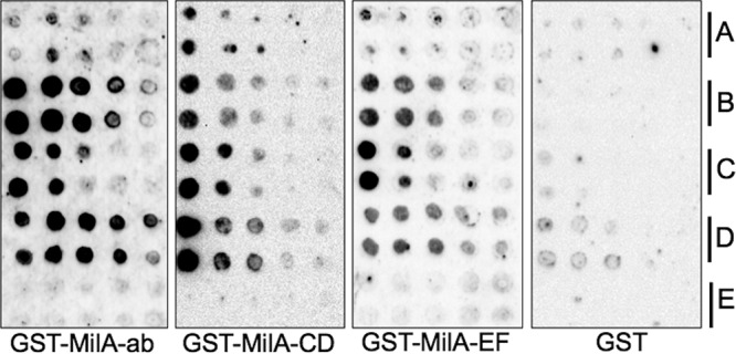
The survival, replication, and virulence of mycoplasmas depend on their ability to capture and import host-derived nutrients using poorly characterized membrane proteins. Previous studies on the important bovine pathogen Mycoplasma bovis demonstrated that the amino-terminal end of an immunogenic 226-kDa (P226) protein, encoded by milA (the full-length product of which has a predicted molecular weight of 303 kDa), had lipase activity. The predicted sequence of MilA contains glycosaminoglycan binding motifs, as well as multiple copies of a domain of unknown function (DUF445) that is also found in apolipoproteins. We mutagenized the gene to facilitate expression of a series of regions spanning the gene in Escherichia coli Using monospecific antibodies against these recombinant proteins, we showed that MilA was proteolytically processed into 226-kDa and 50-kDa fragments that were both partitioned into the detergent phase by Triton X-114 phase fractionation. Trypsin treatment of intact cells showed that P226 was surface exposed. In vitro, the recombinant regions of MilA bound to 1-anilinonaphthalene-8-sulfonic acid and to a variety of lipids. The MilA fragments were also shown to bind heparin. Antibody against the carboxyl-terminal fragment inhibited the growth of M. bovisin vitro This carboxyl end also bound and hydrolyzed ATP, suggestive of a potential role as an autotransporter. Our studies have demonstrated that DUF445 has lipid binding activity and that MilA is a multifunctional protein that may play multiple roles in the pathogenesis of infection with M. bovis.

Keywords: ATPase activity; GAG; Mycoplasma bovis; immunogenicity; lipid binding; membrane protein.


Figures

FIG 1
FIG 1
Physical map of M. bovis PG45 milA. (A) The milA gene lies downstream of rpoB and rpoC and is followed by a pseudogene and an ISMbov6 transposase gene encoded on the opposite strand. The numbers below the genes indicate the number of amino acids encoded by the open reading frame (ORF). (B) Motifs and domains identified in milA, which include the GDSL lipase motif, a recurring domain of unknown function (DUF445) and an unassigned conserved protein domain, COG5283. (C) The recombinant regions expressed during this study. The numbers below the black rectangles indicate the numbers of amino acids in the recombinant MilA protein fragments.
FIG 2
FIG 2
Proteolytic processing of MilA. Proteins in trypsin-treated whole M. bovis PG45 cells, uninoculated M. bovis broth, and the supernatant from a M. bovis broth culture were resolved by SDS-4 to 20% gradient PAGE and either (A) stained with Coomassie blue or (B) transferred onto a polyvinylidene difluoride (PVDF) membrane and probed with a pool of antisera raised in rats against recombinant MilA fragments spanning the whole length of the gene. In addition, whole M. bovis cells were either not treated (NT), or were treated with increasing concentrations of trypsin (0.5, 1, 4, 8, 16, or 32 μg/ml), then their proteins were resolved by SDS-PAGE, transferred onto a PVDF membrane, and probed with rat antisera against MilA-ab (C) or MilA-EF (D). Antibody binding was detected using the Clarity Western ECL blotting substrate (Bio-Rad). (B) The 226- and 50-kDa cleavage fragments of MilA can be seen, with the 226-kDa fragment present in both the mycoplasma cells and the supernatant and susceptible to trypsin cleavage, while the 50-kDa fragment was only detected in the mycoplasma cells and was not cleaved by trypsin. The 226-kDa fragment was detected by antisera against recombinant fragments from the amino and carboxyl ends of the full-length protein (C and D), while the 50-kDa fragment was only detected by the antiserum against the carboxyl end (D). M, PageRuler prestained protein ladder (Thermo Scientific).
FIG 3
FIG 3
Lipid binding properties of M. bovis MilA. Duplicate rows of different lipids at doubling dilutions were dotted onto nitrocellulose membranes. The lipid blots were incubated with purified recombinant MilA-ab, MilA-CD, MilA-EF, or GST (as a negative control). Protein bound to the lipids was detected with goat anti-GST antibody, rabbit anti-goat horseradish peroxidase (HRP) conjugate, and the Clarity Western ECL blotting substrate (Bio-Rad). The lipids on the blots were (A) tributyrin, (B) palmitic acid, (C) oleic acid, (D) cholesterol, and (E) deoxycholic acid.
FIG 4
FIG 4
The binding of M. bovis MilA fragments to the fluorescent probe 1,8-ANS. Recombinant protein fragments spanning MilA (1 μM each) were incubated with 5 μM 1,8-ANS for 3 min at room temperature, and fluorescence was detected by excitation at 385 nm with emission measured at 485 nm in a Hybrid multimode microplate reader (BioTek). The amount of fluorescence (relative fluorescence units [RFU]) corresponds to the amount of 1,8-ANS bound to protein. Data are presented as the mean ± standard deviation (SD); ****, significant differences at P < 0.0001; ***, P = 0.0002; **, P = 0.0026.
FIG 5
FIG 5
Heparin binding by different regions of MilA. (A) Coomassie blue-stained SDS-PAGE of MilA. (B) A duplicate blot of that shown in panel A after Western transfer and probing with biotinylated heparin (100 μg/ml). Binding was detected with goat anti-biotin HRP conjugate (Cell Signaling) and the Clarity Western ECL blotting substrate (Bio-Rad). GST and bovine serum albumin (BSA) served as a negative controls, while GST-0232 served as a positive control (unpublished data). (C) Specificity of heparin binding by MilA-ab. Protein was transferred onto PVDF membrane, blocked, cut into strips, and then treated with increasing amounts of unlabeled heparin or monospecific anti-MilA-ab before probing with biotinylated heparin (100 μg/ml). Binding was detected with goat anti-biotin HRP conjugate (Cell Signaling) and the Clarity Western ECL blotting substrate (Bio-Rad).
FIG 6
FIG 6
Heparin affinity binding and effect of denaturation on binding of MilA to heparin. (A) Doubling dilutions of each of the proteins were blotted onto duplicate nitrocellulose membranes. Blots were stained with Coomassie blue (panel 1) or probed with biotinylated heparin (100 μg/ml) (panel 2). Binding was detected with goat anti-biotin HRP conjugate and ECL substrate (GE Healthcare). (B). Equimolar amounts of recombinant proteins in their native form and after denaturation by heating in cracking buffer were blotted onto nitrocellulose membrane, with GST-MilA-ab in column 1 and GST-MilA-EF in column 2. The first blot was stained with Coomassie blue, while the other blots were incubated in biotinylated heparin (100 μg/ml). Binding was detected with goat anti-biotin HRP conjugate and the ECL Chemiluminescence Substrate (GE Healthcare).
FIG 7
FIG 7
Effect of ATP on tryptic digestion of MilA. Following tryptic digestion of 1 μg of recombinant MilA-EF in the presence or absence of 5 mM ATP and in the presence or absence of 5 mM MgCl2, the fragments were resolved by SDS-12% PAGE and detected by Western immunoblotting, using antisera raised against MilA-EF to probe the blot. (A) MilA-EF stained with Coomassie blue. (B) Western blot of tryptic fragments generated in the absence of MgCl2. (C) Western blot of tryptic digestion products generated in the presence of MgCl2. Lane 1, protein treated with trypsin only; lane 2, protein treated with trypsin in the presence of ATP; lane 3, protein treated with EDTA as a chelator prior to trypsin treatment. M, PageRuler prestained protein ladder (Thermo Scientific).
FIG 8
FIG 8
ATP hydrolysis by MilA. Recombinant protein (60 pmol) was incubated with ATP (1 mM) for 50 min at room temperature and then the malachite green reagent was added and the reaction mixtures were incubated for a further 50 min at room temperature to detect free phosphate. Absorbance was read at 620 nm. BSA and GST were used as negative-control proteins, while inorganic phosphate was used as a positive control. Data are presented as the mean ± SD; ***, P = 0.0002.

Similar articles

Cited by

References

    1. Nicholas R. 2011. Bovine mycoplasmosis: silent and deadly. Vet Rec 168:459–462. doi:10.1136/vr.d2468. - DOI - PubMed
    1. Arcangioli MA, Duet A, Meyer G, Dernburg A, Bezille P, Poumarat F, Le Grand D. 2008. The role of Mycoplasma bovis in bovine respiratory disease outbreaks in veal calf feedlots. Vet J 177:89–93. doi:10.1016/j.tvjl.2007.03.008. - DOI - PubMed
    1. O’Brien D, Scudamore J, Charlier J, Delavergne M. 2016. DISCONTOOLS: a database to identify research gaps on vaccines, pharmaceuticals and diagnostics for the control of infectious diseases of animals. BMC Vet Res 13:1. doi:10.1186/s12917-016-0931-1. - DOI - PMC - PubMed
    1. Buchenau I, Poumarat F, Le Grand D, Linkner H, Rosengarten R, Hewicker-Trautwein M. 2010. Expression of Mycoplasma bovis variable surface membrane proteins in the respiratory tract of calves after experimental infection with a clonal variant of Mycoplasma bovis type strain PG45. Res Vet Sci 89:223–229. doi:10.1016/j.rvsc.2010.03.014. - DOI - PubMed
    1. Caswell JL, Archambault M. 2007. Mycoplasma bovis pneumonia in cattle. Anim Health Res Rev 8:161–186. doi:10.1017/S1466252307001351. - DOI - PubMed

Publication types

MeSH terms

LinkOut - more resources