Review
. 2020 Jun 22;88(7):e00920-19.
doi: 10.1128/IAI.00920-19. Print 2020 Jun 22.

Outer Membrane Lipid Secretion and the Innate Immune Response to Gram-Negative Bacteria

Affiliations
Review

Outer Membrane Lipid Secretion and the Innate Immune Response to Gram-Negative Bacteria

Nicole P Giordano et al. Infect Immun. .

Abstract

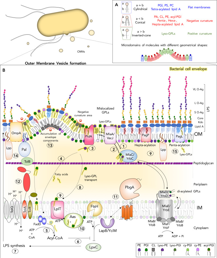
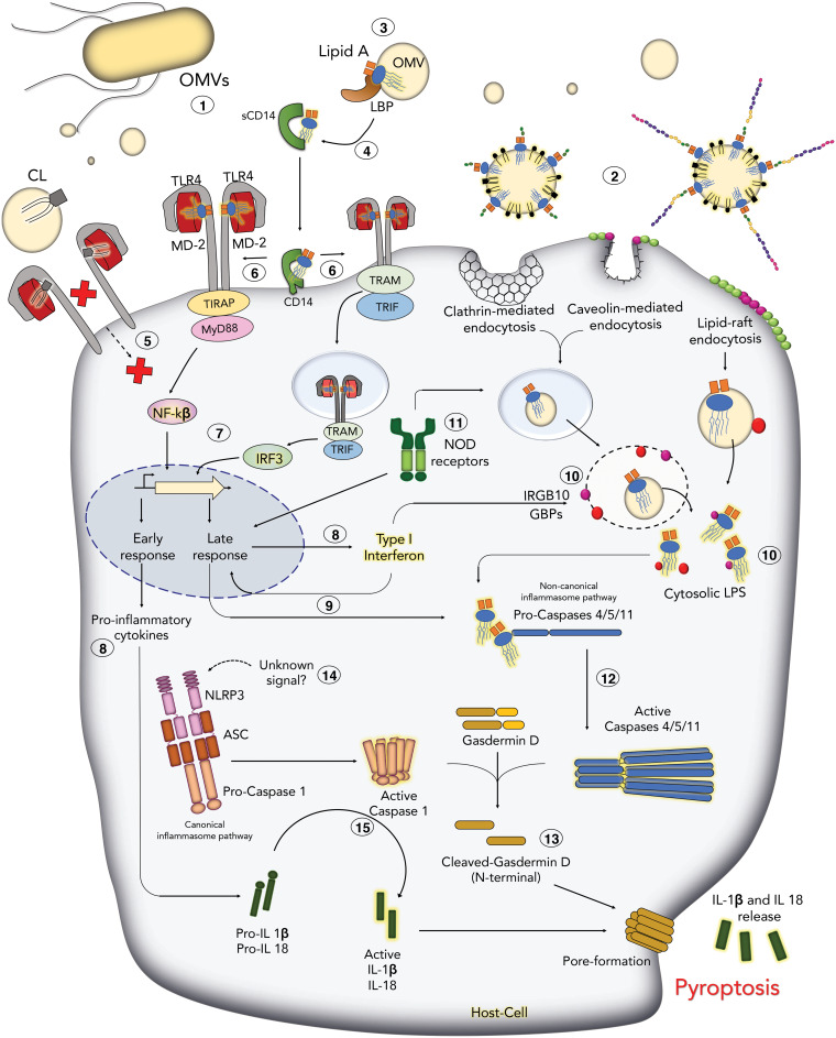
The outer membrane (OM) of Gram-negative bacteria is an asymmetric lipid bilayer that consists of inner leaflet phospholipids and outer leaflet lipopolysaccharides (LPS). The asymmetric character and unique biochemistry of LPS molecules contribute to the OM's ability to function as a molecular permeability barrier that protects the bacterium against hazards in the environment. Assembly and regulation of the OM have been extensively studied for understanding mechanisms of antibiotic resistance and bacterial defense against host immunity; however, there is little knowledge on how Gram-negative bacteria release their OMs into their environment to manipulate their hosts. Discoveries in bacterial lipid trafficking, OM lipid homeostasis, and host recognition of microbial patterns have shed new light on how microbes secrete OM vesicles (OMVs) to influence inflammation, cell death, and disease pathogenesis. Pathogens release OMVs that contain phospholipids, like cardiolipins, and components of LPS molecules, like lipid A endotoxins. These multiacylated lipid amphiphiles are molecular patterns that are differentially detected by host receptors like the Toll-like receptor 4/myeloid differentiation factor 2 complex (TLR4/MD-2), mouse caspase-11, and human caspases 4 and 5. We discuss how lipid ligands on OMVs engage these pattern recognition receptors on the membranes and in the cytosol of mammalian cells. We then detail how bacteria regulate OM lipid asymmetry, negative membrane curvature, and the phospholipid-to-LPS ratio to control OMV formation. The goal is to highlight intersections between OM lipid regulation and host immunity and to provide working models for how bacterial lipids influence vesicle formation.

Keywords: 4; MD-2; OMV; TLR4; Tol-Pal; Toll-like receptor; and 5; antibiotic resistance; antibiotics; antimicrobial peptides; asymmetry; bleb; cardiolipin; caspases 11; constriction; endocytosis; endotoxin; ftsH; gasdermin; glycerophospholipid; immunity; inflammasome; inflammation; lapB/yciM; lipid A; lipid rafts; lipooligosaccharides; lipopolysaccharide; lpp; lpxC; microbial associated molecular patterns; mla; myeloid differentiation factor 2; ompA; outer membrane vesicles; pagL; pagP; pattern recognition receptor; pbgA; peptidoglycan; permeability barrier; pldA; pyroptosis; secretion systems; yejM.


Figures

FIG 1
FIG 1
Bacterial lipid trafficking, remodeling, and homeostasis systems involved in outer membrane vesicle (OMV) biogenesis. Gram-negative bacteria are diderm organisms whose cell envelope consists of an inner membrane (IM) and an outer membrane (OM) that are separated by a periplasmic space and a peptidoglycan layer. Lipid A is the amphipathic constituent of lipopolysaccharide (LPS) and forms the outer leaflet of the OM, while the inner leaflet consists of glycerophospholipids (GPL), making it an asymmetric bilayer. Gram-negative bacteria secrete lipids from their OMs in the form of OMVs and a variety of protein, lipid, and glycolipid components of the cell envelope. The process involves increasing the levels of amphipathic molecules in the outer leaflet of the OM, while not correspondingly increasing the levels in the inner leaflet. Depending upon the shape of lipids that accumulate in these microdomains, the bilayer can curve and pinch off from the surface. (A) Based upon a cross-sectional analysis of the head groups versus the acyl chains, GPLs and lipid A molecules adopt different geometrical shapes, such as conical, inverted conical, or cylindrical, which facilitates negative, positive, or lack of membrane curvature, respectively. (B) Gram-negative bacteria harbor multiple protein systems that work to maintain OM lipid asymmetry. (Step 1) The OM lipoprotein MlaA/VacJ prevents diffusion of GPLs into the OM outer leaflet by adopting an integral membrane conformation. (Step 2) The model of retrograde trafficking for Mla posits that MlaA donates the mislocalized GPLs to MlaC, and MlaC is an acceptor of phosphatidyglycerols (PGl) and phosphatidylethanolamines (PE). MlaC would bind to the MlaFEDB/YrbFEDB complex in the IM and transfer the GPLs to MlaD for their reintegration into the IM. The anterograde model posits that MlaC donates GPLs to MlaA, which is an acceptor in the OM. An anterograde mechanism would involve MlaC receiving GPLs from the IM complex and transporting them outwardly across the periplasm to the OM. In the OM, GPLs flip into the outer leaflet at a low frequency during growth and a greater frequency during OM damage. (Step 3) OM outer leaflet mislocalized GPLs are substrates for PldA, a beta-barrel phospholipase that generates lysophospholipids and fatty acids in the outer leaflet to maintain asymmetry (Step 4). Fatty acids diffuse back across the periplasm, cross the IM, and are converted by FadD to acyl-CoA (Step 5). Acyl-CoA, or another product originated from phospholipid metabolism in the OM, might act as a second messenger to modulate LpxC degradation (Step 6). This would allow bacteria to pause LpxC degradation and increase LPS biosynthesis (Step 7), thereby restoring balance to the level of GPLs and glycolipids that comprise the OM. PldA, and the OM beta barrel enzyme PagP, generate lysophospholipids (lyso-GPLs) from inverted GPLs. (Step 8) Lysophospholipids can act as detergents that disrupt the bilayer and bacteria presumably traffic these molecules back to the IM; however, this mechanism is not known. (Step 9) On their return to the IM, lysophospholipids are flipped across the membrane by the flippase, LplT. (Step 10) On the cytoplasmic surface, the acyltransferase Aas restores the lysophospholipids to their diacyl form. (Step 11) PbgA/YejM is an essential lipid-binding protein involved in regulating LPS biosynthesis. The model posits that PbgA-mediated LPS regulation works through LapB/YciM and LapB’s ability to modulate FtsH-mediated protein digestion, especially for LpxC. (Step 12) The Tol-Pal system forms a trans-envelope protein complex important for constricting the OM by forming energized interactions between the OM lipoprotein Pal and peptidoglycan, promoting-negative membrane curvature, and maintaining OM integrity. Loss of Tol-Pal causes GPLs to accumulate in the OM and results in OMV formation. (Step 13) Peptidoglycan fragments and misfolded proteins can also increase in the periplasm and cause changes in turgor pressure between the peptidoglycan layer and OM that might cause OMVs to form. (Step 14) In addition, major OM proteins like OmpA and the OM lipoprotein Lpp form periplasmic contacts with peptidoglycan, and alterations in these interactions have been shown to result in OMV release. (Step 15) Lipid A modifications by the PagP palmitoyltransferase and the PagL demyristoylase result in hyper- or hypoacylated forms of lipid A for LPS molecules, respectively. These changes in the lipid A acylation state impact the shape of the molecules in the outer leaflet and perhaps influence the local curvature of the bilayer. Abbreviations: CL, cardiolipin; PA, phosphatidic acid; PS, phosphatidylserine; acyl-PGL, acyl-phosphatidylglycerol; PC, phosphorylcholine; S O-Ag, short O-antigen; L O-Ag, long O-antigen; VL O-Ag, very long O-antigen; ATP, adenosine triphosphate.
FIG 2
FIG 2
Innate immune pattern recognition of bacterial lipids on OMVs leads to inflammation and death. Lipid A and cardiolipin (CL) are constituents of OMVs that engage pattern recognition receptors on membranes and in the cytosol of host cells. The LPS molecules carried by OMVs interact with a variety of proteins and systems. Lipid A components of these LPS molecules activate proinflammatory cytokine production and trigger an inflammatory cell death known as pyroptosis. Once released, OMVs (Step 1) can enter host cells through endocytosis (Step 2). In the bloodstream, OMVs interact with the circulating plasma protein, LPS-binding protein, or LBP (Step 3), which extracts a single LPS molecule from the micelle. LBP binds to soluble CD14 (sCD14) and membrane-anchored CD14 molecules. Interactions with LBP, CD14, and LPS result in the remodeling and presentation of the lipid A disaccharolipid component of LPS to MD-2 (Step 4). MD-2 interacts with lipid A by a sandwich-like mechanism, which influences TLR4 receptor dimerization and activation. CL can also interact with the TLR4/MD-2 complex on the surface (Step 5) and can act as an agonist or antagonist. Dimerization of TLR4/MD-2 (Step 6) recruits different adaptor proteins that will activate transcription factors such as nuclear factor κβ (NFκβ) and interferon regulatory factor 3 (IRF3) (Step 7), which will ultimately result in production of proinflammatory cytokines (inactive pro-IL-1β and pro-IL-18) and type I interferon (IFN-β) (Step 8). Type-I IFN also induces the expression of caspase-11 (Step 9). The production of type I IFN after TLR4 activation enhances the expression of cytoplasmic proteins, such as Interferon-related GTPase-10 (IRGB10) and guanylate-binding proteins (GBPs), which bind pathogen vacuoles and LPS molecules on cytosolic bacteria and OMVs (Step 10), and might facilitate endosome lysis or release of LPS molecules, and ultimately lipid A endotoxins, for detection by caspases-4/5/11. NOD receptors bind peptidoglycan fragments on OMVs and activate late inflammatory responses to pathogens (Step 11). Binding of the lipid A disaccharolipid by caspases-4/5/11 promotes autoprocessing and activation (Step 12). Activated caspase-4/5/11 cleaves gasdermin D, which causes gasdermin D to oligomerize and form large pores in the plasma membrane that alter ion homeostasis and cause cell lysis (Step 13). The NLRP3 noncanonical inflammasome is activated by an unknown signal that is downstream caspase-4/5/11 activation by lipid A molecules (Step 14). Signals received by NLRP3 activate autoprocessing of caspase-1 and activation of the canonical inflammasome. Active caspase-1 cleaves IL-1β and IL-18, as well as gasdermin D, which result in robust proinflammatory cell death, or pyroptosis (Step 15). The dual activation pathway combines the canonical and noncanonical inflammasomes and is specific to innate immune cells, which we have attempted to depict here.

References

    1. Silhavy TJ, Kahne D, Walker S. 2010. The bacterial cell envelope. Cold Spring Harb Perspect Biol 2:a000414. doi: 10.1101/cshperspect.a000414. - DOI - PMC - PubMed
    1. Powers MJ, Trent MS. 2019. Intermembrane transport: glycerophospholipid homeostasis of the Gram-negative cell envelope. Proc Natl Acad Sci U S A 116:17147–17155. doi: 10.1073/pnas.1902026116. - DOI - PMC - PubMed
    1. Shrivastava R, Chng SS. 2019. Lipid trafficking across the Gram-negative cell envelope. J Biol Chem 294:14175–14184. doi: 10.1074/jbc.AW119.008139. - DOI - PMC - PubMed
    1. Simpson BW, Trent MS. 2019. Pushing the envelope: LPS modifications and their consequences. Nat Rev Microbiol 17:403–416. doi: 10.1038/s41579-019-0201-x. - DOI - PMC - PubMed
    1. Gioannini TL, Teghanemt A, Zhang D, Coussens NP, Dockstader W, Ramaswamy S, Weiss JP. 2004. Isolation of an endotoxin-MD-2 complex that produces Toll-like receptor 4-dependent cell activation at picomolar concentrations. Proc Natl Acad Sci U S A 101:4186–4191. doi: 10.1073/pnas.0306906101. - DOI - PMC - PubMed

Publication types

MeSH terms

LinkOut - more resources