. 2020 Jun;58(6):1309-1323.
doi: 10.1007/s11517-020-02142-8. Epub 2020 Apr 7.

An optimisation-based iterative approach for speckle tracking echocardiography

Affiliations

An optimisation-based iterative approach for speckle tracking echocardiography

Neda Azarmehr et al. Med Biol Eng Comput. 2020 Jun.

Abstract

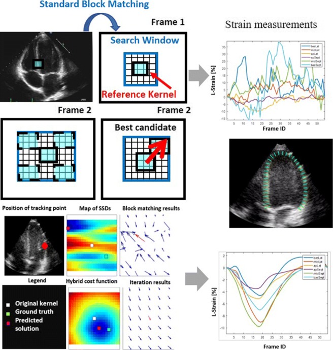
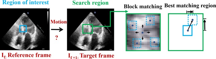
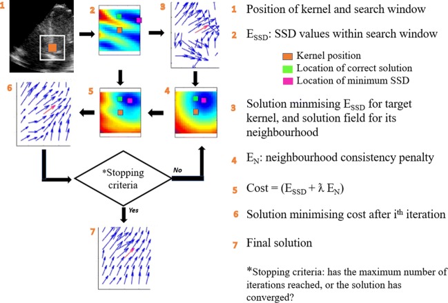
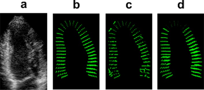
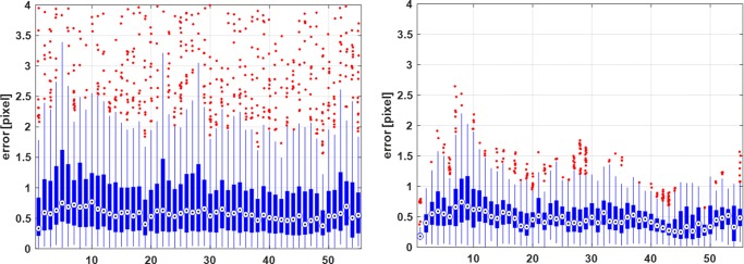
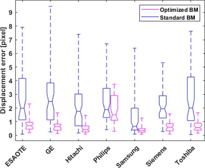
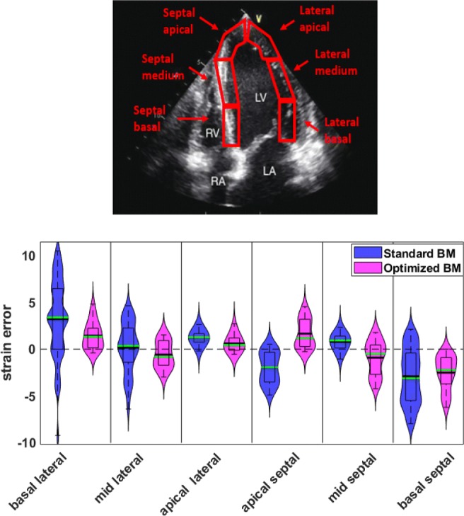
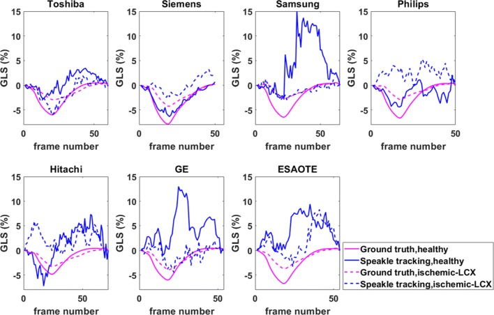
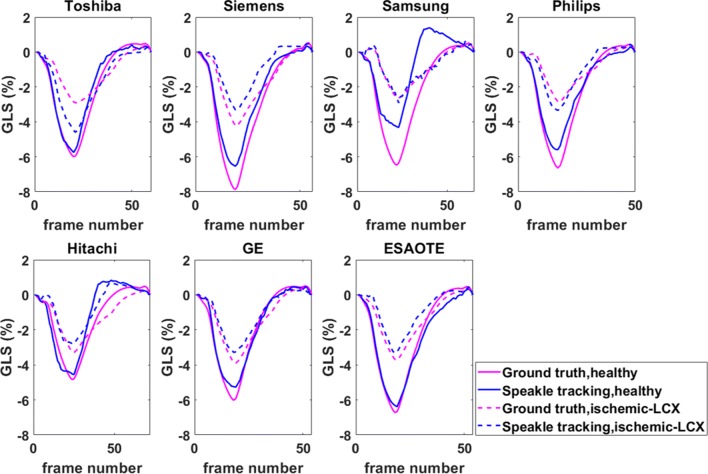
Speckle tracking is the most prominent technique used to estimate the regional movement of the heart based on echocardiograms. In this study, we propose an optimised-based block matching algorithm to perform speckle tracking iteratively. The proposed technique was evaluated using a publicly available synthetic echocardiographic dataset with known ground-truth from several major vendors and for healthy/ischaemic cases. The results were compared with the results from the classic (standard) two-dimensional block matching. The proposed method presented an average displacement error of 0.57 pixels, while classic block matching provided an average error of 1.15 pixels. When estimating the segmental/regional longitudinal strain in healthy cases, the proposed method, with an average of 0.32 ± 0.53, outperformed the classic counterpart, with an average of 3.43 ± 2.84. A similar superior performance was observed in ischaemic cases. This method does not require any additional ad hoc filtering process. Therefore, it can potentially help to reduce the variability in the strain measurements caused by various post-processing techniques applied by different implementations of the speckle tracking. Graphical Abstract Standard block matching versus proposed iterative block matching approach.

Keywords: Echocardiography; Myocardial deformation; Speckle tracking echocardiography; Strain imaging.


Figures

Graphical Abstract
Graphical Abstract
Standard block matching versus proposed iterative block matching approach
Fig. 1
Fig. 1
Speckle tracking using BM where a region in the image (kernel) is selected and sought for in the next image by sequentially trying out different positions, testing the similarity between the kernel and the pattern observed in that position. The position where the similarity between the kernel and the observed pattern is maximal is accepted as the new position of the original kernel
Fig. 2
Fig. 2
Flowchart showing the steps involved in solving the proposed optimisation-based tracking algorithm
Fig. 3
Fig. 3
An example A4C from the Siemens healthy sequence and corresponding displacement vector fields during the rapid ejection phase (peak systole). a Zoomed view of LV cropped from the original image. b Ground-truth. cd Displacement fields obtained from standard BM and optimised BM approach in the rapid ejection phase, respectively. Corresponding figures for other vendors are provided in Appendix A
Fig. 4
Fig. 4
Boxplots of the error for the healthy sequence from Siemens. The error is computed as the magnitude of the difference between the calculated and ground-truth displacement vectors and is provided for standard (left) and proposed (right) tracking methods. The x-axis shows the frame number
Fig. 5
Fig. 5
Displacement error for the healthy A4C synthetic sequences across all vendors for the two speckle tracking approaches. The horizontal line represents mean; the box signifies the quartiles, and the whiskers represent the 2.5% and 97.5% percentiles
Fig. 6
Fig. 6
Top: an A4C view with the LV myocardium segmentation regions overlaid. Below: violin plots of the error in the segmental strain measurements for the healthy synthetic sequence from Siemens. The solid black line represents mean, and the green line represents the median; the box signifies the quartiles, and the whiskers represent the 2.5% and 97.5% percentiles
Fig. 7
Fig. 7
Comparison of GLS measurements obtained from the standard BM approach for the healthy and ischaemic LCX (ischaemic left circumflex coronary artery) cases across all vendors with the known ground-truth. The solid and dashed blue lines represent the calculated strain values for healthy and ischaemic cases, respectively. The solid and dashed magenta lines indicate the corresponding ground-truth
Fig. 8
Fig. 8
Same as Fig. 7, but for the optimised BM approach
Fig. 9
Fig. 9
Example of a (presumably) “common sense” editing in a commercial package on one frame, where 3 regional strain values are given for the lateral wall, but there is no visible myocardium to be tracked in that region

Similar articles

Cited by

References

    1. Voigt JU, Pedrizzetti G, Lysyansky P, Marwick TH, Houle H, Baumann R, Pedri S, Ito Y, Abe Y, Metz S, Song JH. Definitions for a common standard for 2D speckle tracking echocardiography: consensus document of the EACVI/ASE/Industry Task Force to standardize deformation imaging. Eur Heart J Cardiovasc Imaging. 2014;16(1):1–11. doi: 10.1093/ehjci/jeu184. - DOI - PubMed
    1. Kalam K, Otahal P, Marwick TH. Prognostic implications of global LV dysfunction: a systematic review and meta-analysis of global longitudinal stran and ejection fraction. Heart. 2014;100:1673–1680. doi: 10.1136/heartjnl-2014-305538. - DOI - PubMed
    1. Barbosa D, Friboulet D, D’hooge J, Bernard O (2014) Fast tracking of the left ventricle using global anatomical affine optical flow and local recursive block matching. MIDAS J: 10
    1. Ferraiuoli P, Fixsen LS, Kappler B, Lopata RG, Fenner JW, Narracott AJ. Measurement of in vitro cardiac deformation by means of 3D digital image correlation and ultrasound 2D speckle-tracking echocardiography. Medical Engineering & Physics. 2019;74:146–152. doi: 10.1016/j.medengphy.2019.09.021. - DOI - PubMed
    1. Rodriguez A, Fernandez-Lozano C, Dorado J, Rabunal JR. Two-dimensional gel electrophoresis image registration using block-matching techniques and deformation models. Analytical Biochemistry. 2014;454:53–59. doi: 10.1016/j.ab.2014.02.027. - DOI - PubMed

MeSH terms