. 2020 Jun;57(6):2600-2619.
doi: 10.1007/s12035-020-01904-7. Epub 2020 Apr 6.

cGAS/STING Pathway Activation Contributes to Delayed Neurodegeneration in Neonatal Hypoxia-Ischemia Rat Model: Possible Involvement of LINE-1

Affiliations

cGAS/STING Pathway Activation Contributes to Delayed Neurodegeneration in Neonatal Hypoxia-Ischemia Rat Model: Possible Involvement of LINE-1

Marcin Gamdzyk et al. Mol Neurobiol. 2020 Jun.

Abstract

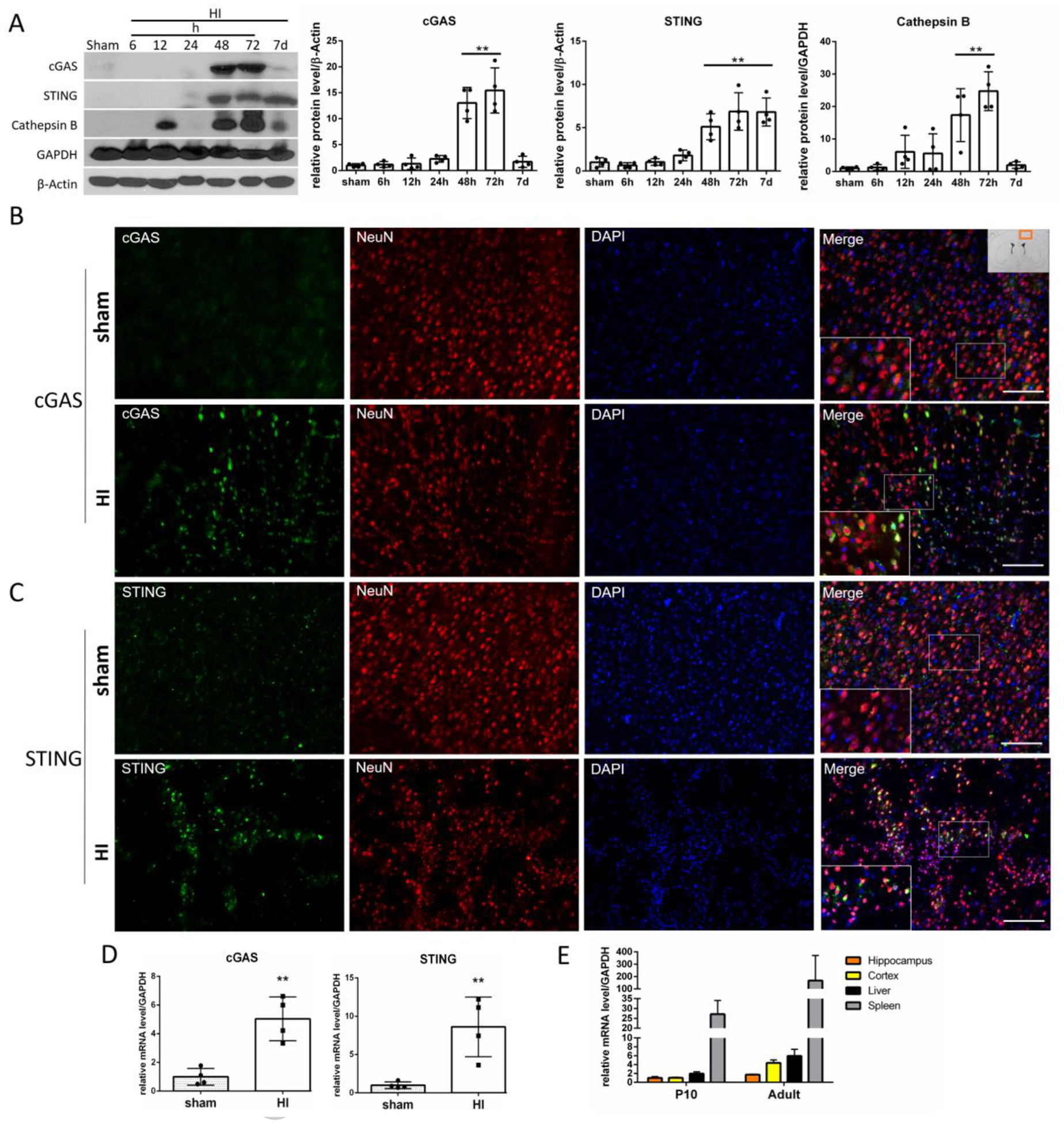
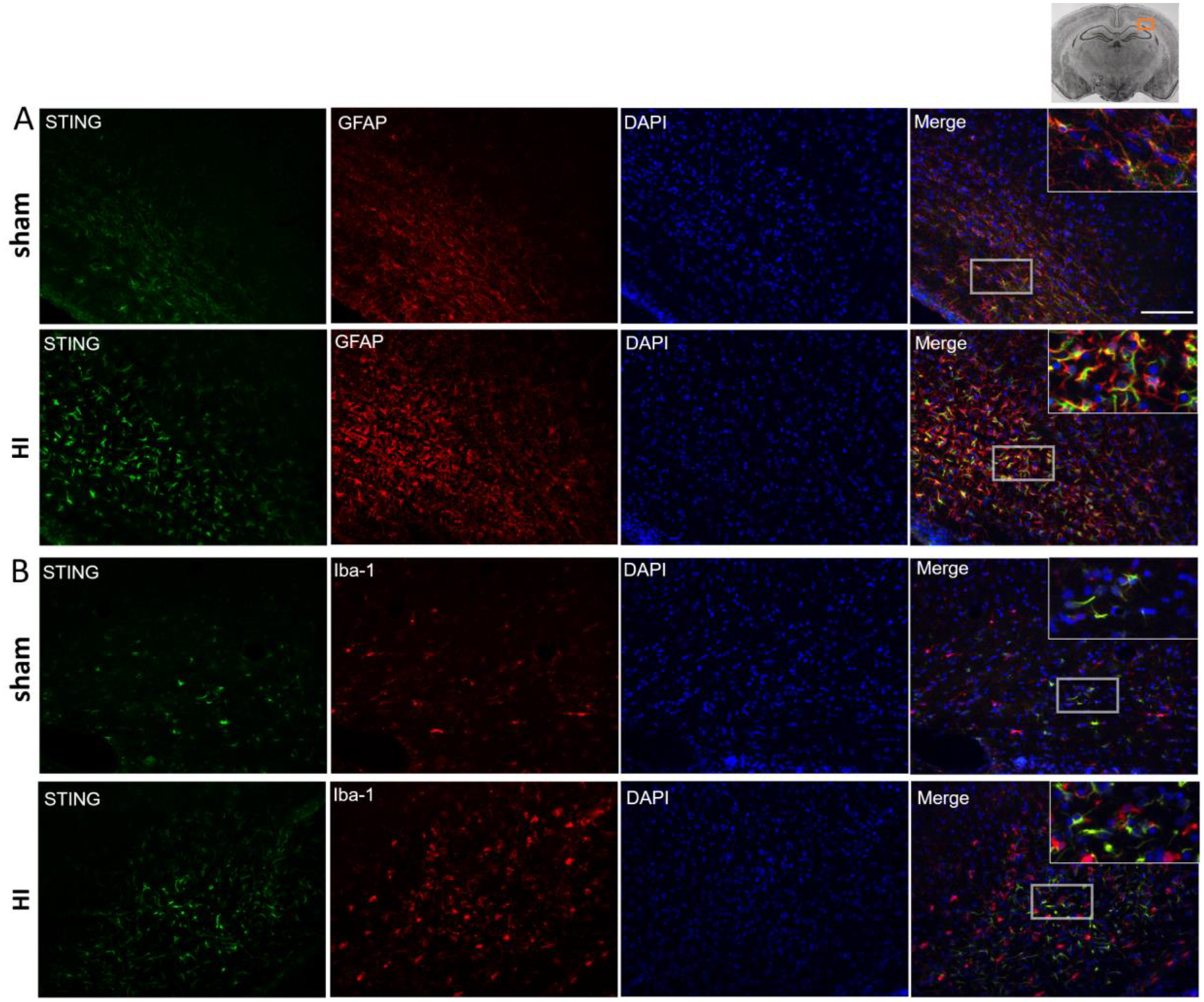
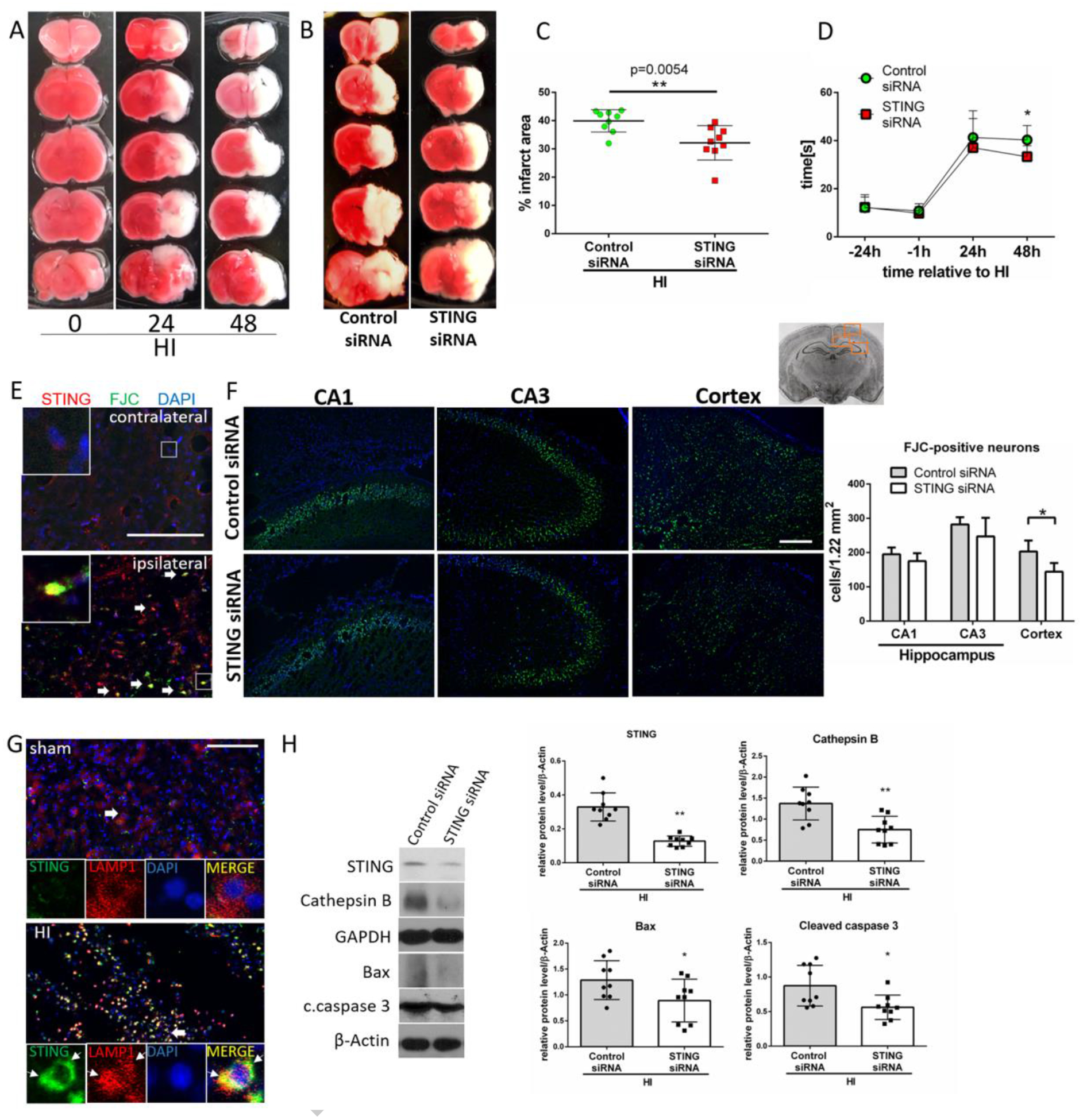
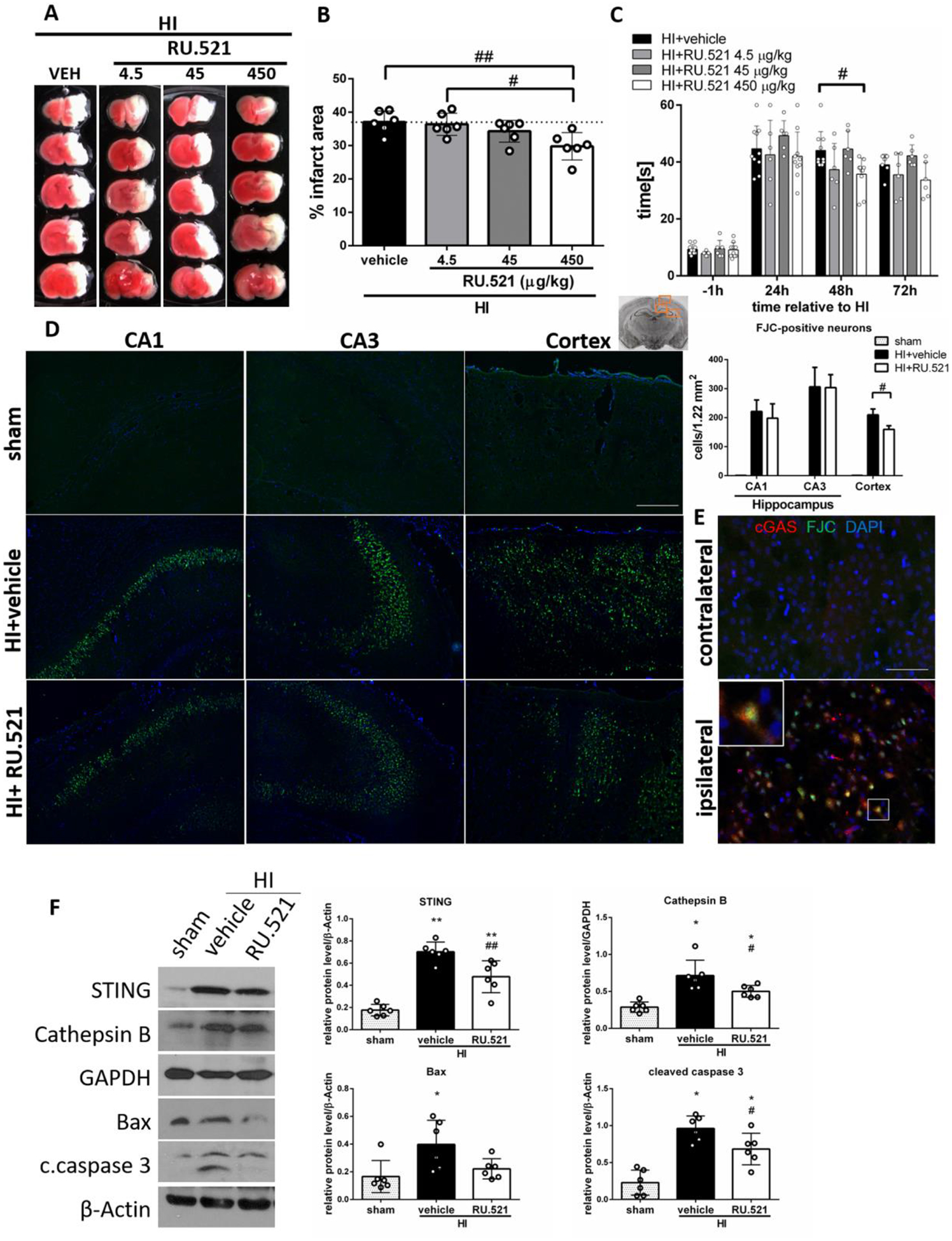
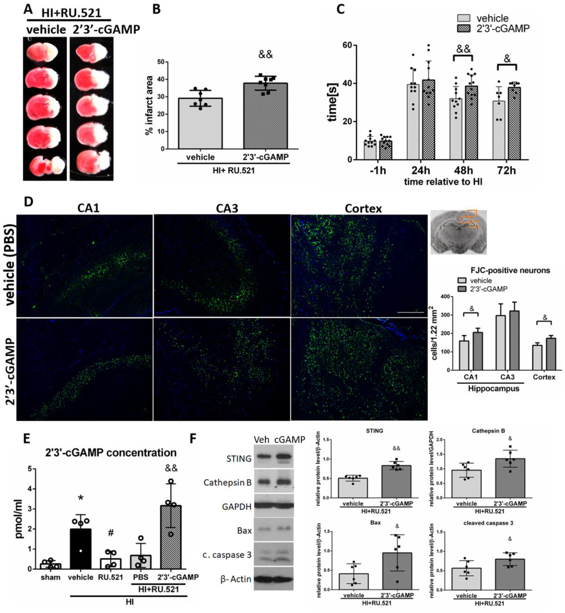
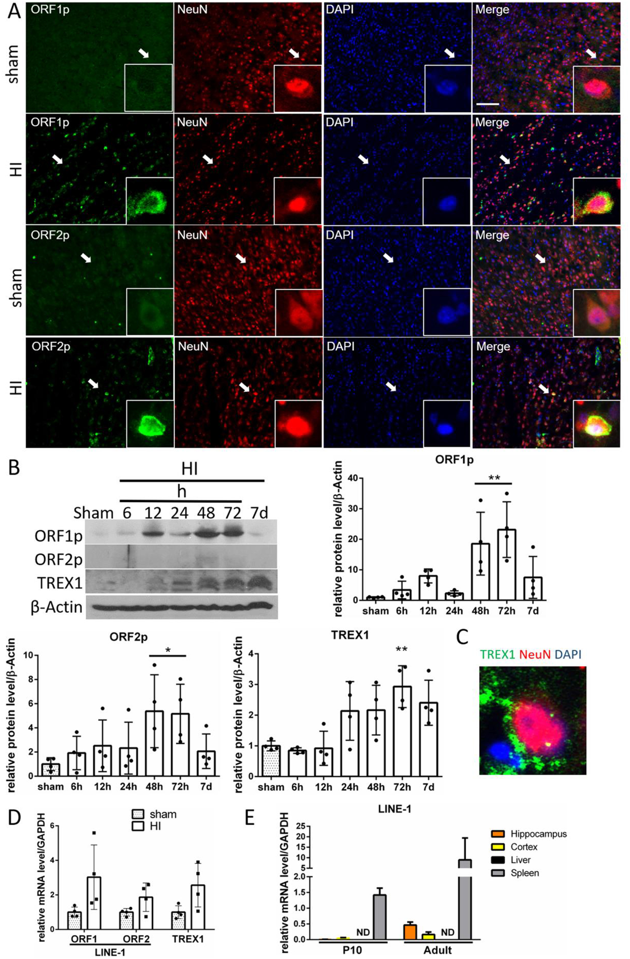
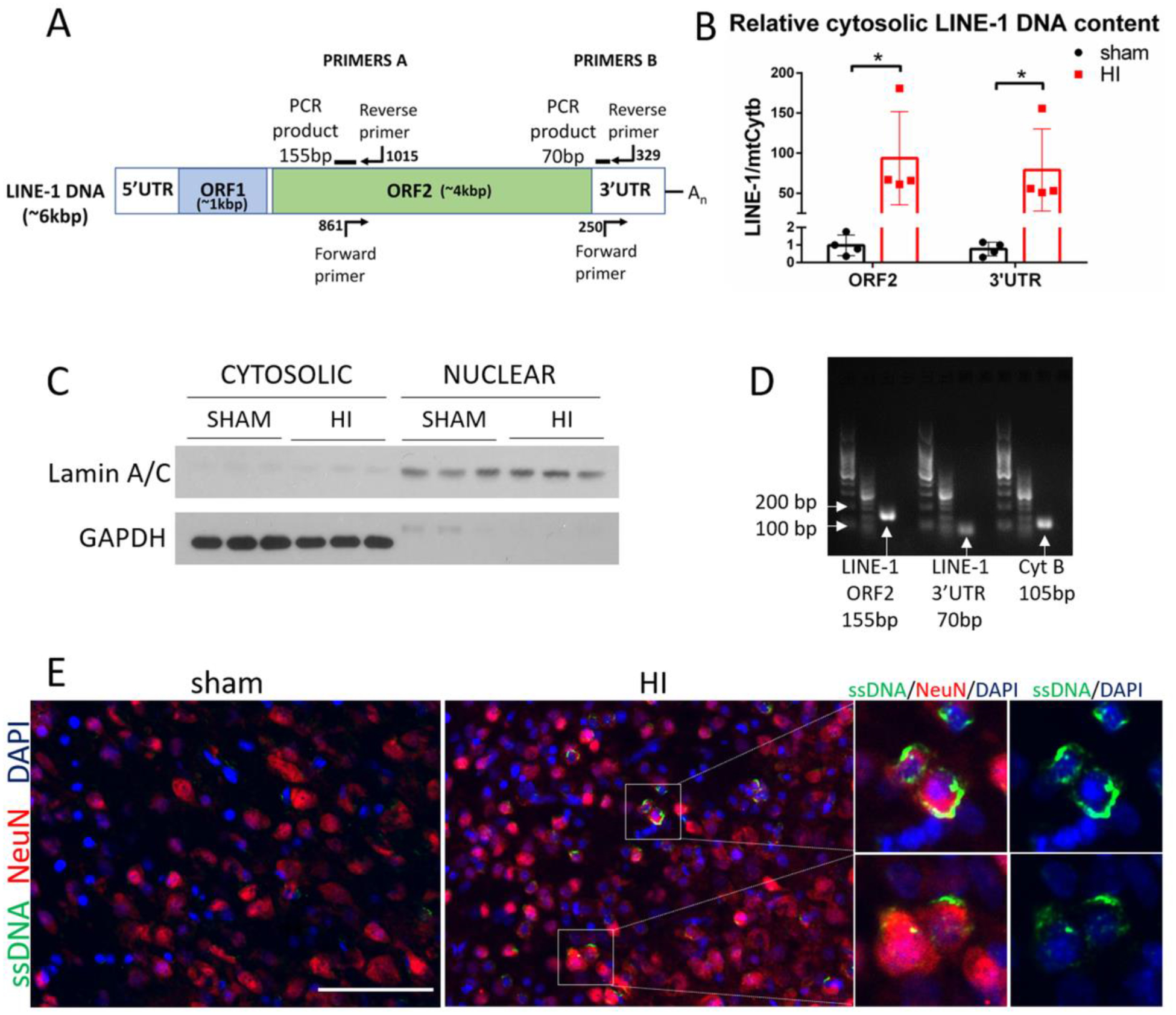
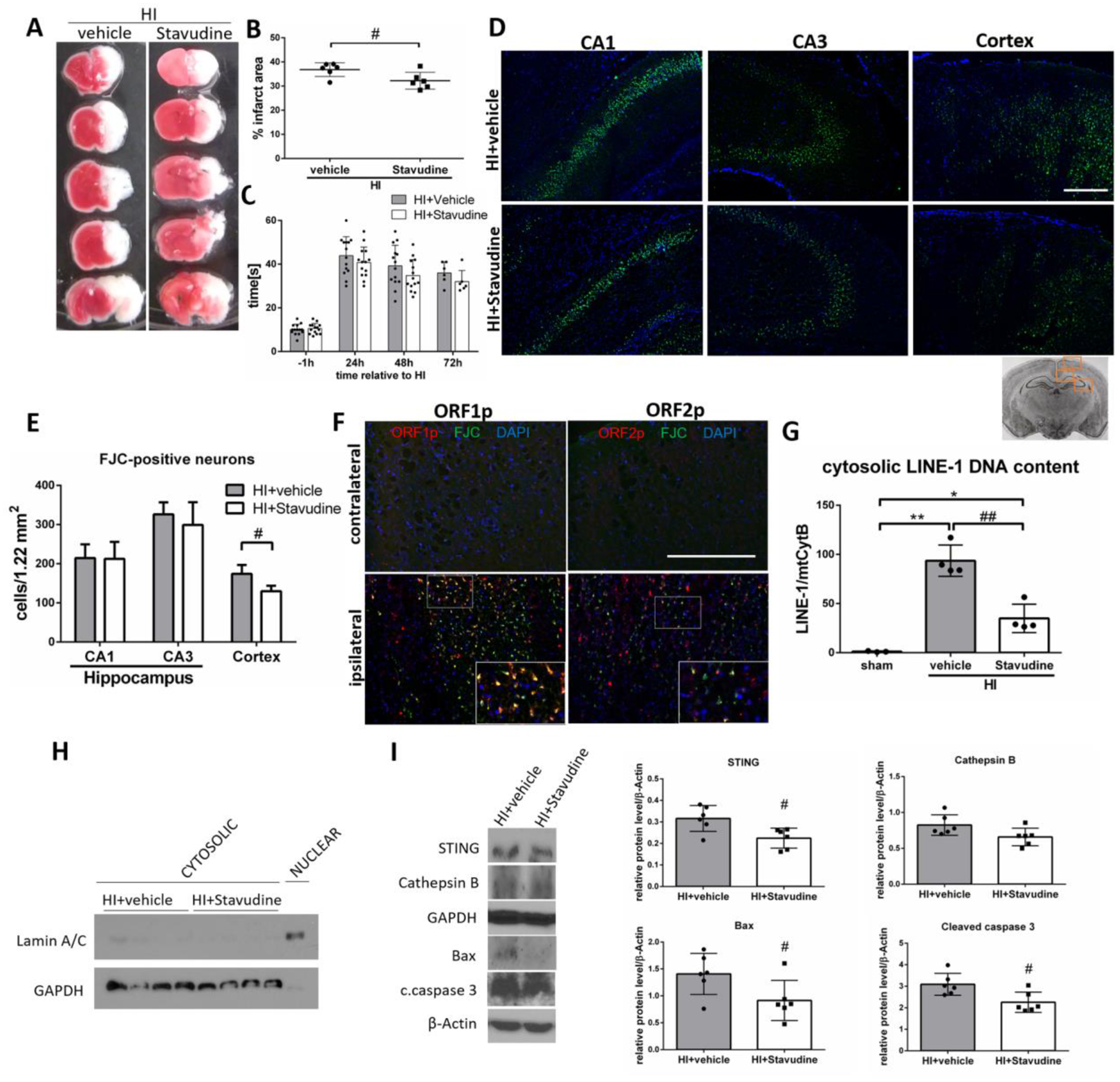
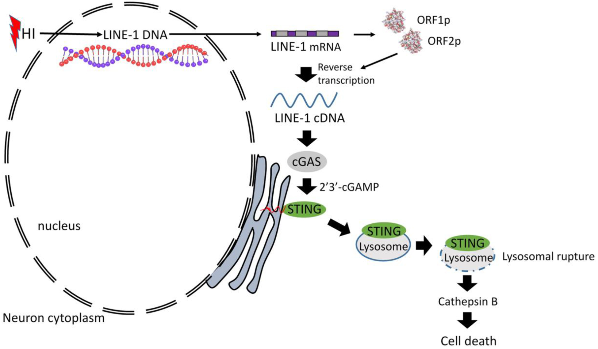
cGAS is a sensor of cytosolic DNA and responds equally to exogenous and endogenous DNA. After recognition of cytosolic dsDNA or ssDNA, cGAS synthesizes the second messenger 2'3'-cGAMP, which then binds to and activates stimulator of interferon genes (STING). STING plays an essential role in responding to pathogenic DNA and self-DNA in the context of autoimmunity. In pathologic conditions, such as stroke or hypoxia-ischemia (HI), DNA can gain access into the cytoplasm of the cell and leak from the dying cells into the extracellular environment, which potentially activates cGAS/STING. Recent in vivo studies of myocardial ischemia, traumatic brain injury, and liver damage models suggest that activation of cGAS/STING is not only a side-effect of the injury, but it can also actively contribute to cell death and apoptosis. We found, for the first time, that cGAS/STING pathway becomes activated between 24 and 48 h after HI in a 10-day-old rat model. Silencing STING with siRNA resulted in decreased infarction area, reduced cortical neurodegeneration, and improved neurobehavior at 48 h, suggesting that STING can contribute to injury progression after HI. STING colocalized with lysosomal marker LAMP-1 and blocking STING reduced the expression of cathepsin B and decreased the expression of Bax and caspase 3 cleavage. We observed similar protective effects after intranasal treatment with cGAS inhibitor RU.521, which were reversed by administration of STING agonist 2'3'-cGAMP. Additionally, we showed that long interspersed element 1 (LINE-1) retrotransposon, a potential upstream activator of cGAS/STING pathway was induced at 48 h after HI, which was evidenced by increased expression of ORF1p and ORF2p proteins and increased LINE-1 DNA content in the cytosol. Blocking LINE-1 with the nucleoside analog reverse-transcriptase inhibitor (NRTI) stavudine reduced infarction area, neuronal degeneration in the cerebral cortex, and reduced the expression of Bax and cleaved caspase 3. Thus, our results identify the cGAS/STING pathway as a potential therapeutic target to inhibit delayed neuronal death after HI.

Keywords: Apoptosis; LINE-1; Neurodegeneration; STING; cGAS.


Conflict of interest statement

Declaration of Interest

The authors declare no conflict of interest for this study.

Figures

Fig.1.
Fig.1.
cGAS/STING pathway is activated after HI. (A) Representative western blot images and quantitative analysis of relative temporal expression of endogenous cGAS, STING and Cathepsin B in the ipsilateral hemisphere of the rat brain after hypoxia-ischemia (HI). Expression of cGAS, STING and Cathepsin B was significantly increased (p<0.01) at 48 and 72h after HI compared to sham. STING was additionally increased at 7 days after HI (p<0.01). Values are expressed as mean ± SD. **p < 0.01 vs. sham group. N = 4. Expression of cGAS (B) and STING (C) is shown by double immunofluorescence staining for cGAS/STING (green) in neurons (NeuN, red) in ipsilateral cerebral cortex in sham and HI animals 48h after HI. Scale bar 100 μm. N = 2. Exemplary cells expressing cGAS/STING are indicated with arrows. Picture in the right upper corner shows approximate location from where the images were taken. (D) cGAS and STING mRNA level quantification in ipsilateral hemisphere. cGAS and STING transcript levels were elevated at 72h after HI. Values are expressed as mean ± SD. **p < 0.01 vs. sham group. N = 4. (E) Detection of STING mRNA in rat tissues. STING mRNA levels were detected in P10 and adult rats in hippocampus, cortex, liver and spleen, with highest expression found in spleen. Values are expressed as mean ± SD. N=2–3.
Fig. 2.
Fig. 2.
Immunofluorescence staining of STING in the rat brain in the ipsilateral hemisphere at 48 h post HI. Immunofluorescence staining showed that STING was colocalized with astrocytic marker GFAP (A) and microglia marker Iba-1 in sham and HI group. Green was for STING, Red was for GFAP (A) or Iba-1 (B), Blue was for DAPI. Merge showed the colocalization. Scale bar = 100 μm.
Fig.3.
Fig.3.
STING inhibition has neuroprotective effects in HI rats. (A) Representative images showing infarct propagation between 24 and 48h after HI. (B) Representative images of brain sections from rats administered with control or STING siRNA stained with TTC at 48h after HI. (C) Infarction area quantification. STING siRNA injection at 48h before HI significantly reduced infarction area in ipsilateral hemisphere at 48h after HI compared to control siRNA (p<0.01). N=9. (D) Negative geotaxis test results. Pups injected with STING siRNA showed decreased neurological impairments at 48h after HI (p<0.05). Two-way-ANOVA-Sidak N=13. (E) Representative microphotographs of double immunofluorescence staining for STING (red) and Fluoro-Jade C (FJC, green) in contralateral and ipsilateral hemisphere of HI rats. Exemplary cells double-positive for STING and FJC are indicated with arrows. (F) Representative images of CA1, CA3 and cerebral cortex stained with Fluoro-Jade C/DAPI and quantitative analysis of FJC-positive neurons. Silencing of STING significantly reduced number of FJC-positive neurons in cerebral cortex (p<0.05) at 48h after HI compared to control siRNA group. N=4. (G) Double immunofluorescence staining for STING and lysosomal marker LAMP1 in cerebral cortex of sham and HI rats. STING colocalized with LAMP1 puncta in ipsilateral hemisphere at 48h after HI. (H) Representative western blot images and quantitative analysis of relative expression of STING, Cathepsin B, Bax and cleaved caspase 3. Silencing of STING significantly decreased level of STING, Cathepsin B, Bax and cleaved caspase 3 in ipsilateral hemisphere at 48h after HI compared to control siRNA group. N=6. Values are expressed as mean ± SD. *p<0.05, **p < 0.01 vs. control siRNA. Scale bar 100μm.
Fig.4.
Fig.4.
RU.521 decreases neuronal death after HI. (A) Representative pictures of TTC-stained brain sections of animals administered with intranasal RU.521 in a dose of 4.5, 45, 450 μg/kg or vehicle. (B) Quantitative analysis of infarction area showed in TTC stained brain slices. 450 μg/kg dose of cGAS inhibitor RU.521 significantly decreased infarction area at 72h after HI (p<0.01). N=6. (C) Geotaxis test showed that 450 μg/kg dose of RU.521 decreased the reflex time significantly (p<0.05) compared with the vehicle group at 48h after HI. N=6–10. (D) Representative images of CA1, CA3 and cerebral cortex stained with Fluoro-Jade C/DAPI and quantitative analysis of FJC-positive neurons. RU.521 (450 μg/kg) significantly reduced (p<0.05) the number of FJC-positive neurons in cerebral cortex of ipsilateral hemisphere at 48h after HI. N=4. Scale bar 100μm. (E) Representative microphotographs of double immunofluorescence staining for cGAS (red) and Fluoro-Jade C (green) in contralateral and ipsilateral hemisphere of HI rats. Scale bar 50μm. (F) Representative western blot images and quantitative analysis of relative expression of STING, Cathepsin B, Bax and cleaved caspase 3 at 72h after HI and after RU.521 treatment. N=6. Data presented as mean ± SD. *p<0.05, **p < 0.01 vs. sham, #p<0.05, ##p < 0.01 vs. vehicle.
Fig.5.
Fig.5.
2’3’-cGAMP reverses RU.521-induced protective effects. (A) Representative brain sections of animals administered with PBS (vehicle) or 2’3’-cGAMP stained with TTC at 72h after HI. (B) Quantitative analysis of infarction area showed in TTC stained brain slices. STING agonist 2’3’-cGAMP significantly increased infarction area at 72h after HI compared to vehicle group. N=7–8. (C) Negative geotaxis test results. 2’3’-cGAMP significantly worsened rat performance in negative geotaxis test compared to vehicle group at 48 and 72h after HI. N=11–12. (D) Representative images of CA1, CA3 and cerebral cortex stained with Fluoro-Jade C/DAPI and quantitative analysis of FJC-positive neurons. 2’3’-cGAMP significantly increased the number of FJC-positive neurons in CA1 area of the hippocampus and in cerebral cortex of ipsilateral hemisphere at 48h after HI. N=4. Scale bar 100μm. (E) ELISA analysis of 2’3’-cGAMP concentration in ipsilateral hemispheres of rats at 72h after HI. N=4. (F) Representative western blot images and quantitative analysis of relative expression of STING, Cathepsin B, Bax and cleaved caspase 3 at 72h after HI. N=6. Data presented as mean ± SD. &p<0.05, &&p < 0.01 vs. 2’3’-cGAMP vehicle (PBS). *p<0.05 vs. sham, #p < 0.05 vs. vehicle. Scale bar 100μm.
Fig.6.
Fig.6.
LINE-1 is activated after HI. (A) Expression of ORF1p and ORF2p proteins (green) is shown by double immunofluorescence staining in neurons (NeuN, red) in sham and HI animals in ipsilateral cerebral cortex at 48h after HI. Arrows indicate cells showed in higher magnification. N=2. (B) Representative western blot images of time-course data and quantitative analysis of relative temporal expression of endogenous ORF1p, ORF2p and TREX1 in the ipsilateral hemisphere of the rat brain after HI. Expression of ORF1p and ORF2p was significantly increased compared to sham at 48 and 72h after HI. TREX1 expression was significantly increased at 72h after HI. (C) TREX1 expression was detected in cortical neurons. (D) Relative level of ORF1, ORF2 and TREX1 mRNA at 72h after HI. (E) Detection of LINE-1 ORF1 mRNA in naïve rat tissues. LINE-1 ORF1 mRNA was detected in P10 and adult rats in hippocampus, cortex and spleen, with highest expression found in spleen. Values are expressed as mean ± SD. ND – nondetectable. N=2–3. **p < 0.01, *p < 0.05 vs. sham group. Scale bar 100μm.
Fig.7.
Fig.7.
LINE-1 DNA accumulates in the cytosol after HI. (A) LINE-1 illustration showing fragments targeted with PCR primers. (B) Relative cytosolic LINE-1 DNA content in sham and HI group was quantified by PCR at 48h after HI. MtCytB was used as a reference gene. (C) Validation of cytosolic and nuclear fractionation by western blot. Cytosolic and nuclear extracts were probed with antibodies against Lamin A/C- a nuclear membrane protein, and with GAPDH – cytosolic protein. (D) LINE-1 PCR products were visualized on agarose gel. (E) Immunostaining for ssDNA in sham and HI animals at 48h after HI. N=2. Magnified panel to the right shows merged images for ssDNA and DAPI to illustrate cytosolic location of ssDNA after HI. Scale bar 100μm.
Fig.8.
Fig.8.
Stavudine inhibits LINE-1 and STING and decreases neuronal death in cerebral cortex after HI. (A) Representative pictures of TTC-stained brain sections of animals administered with intranasal Stavudine or vehicle. (B) Quantitative analysis of infarction area showed in TTC stained brain slices. Stavudine in a dose of 500 μg/kg significantly decreased infarction area compared to vehicle at 72h after HI (p<0.05). N=6. (C) Negative geotaxis test was performed at 1h before, and 24,48 and 72h after HI. N=6–14. (D) Representative images of CA1, CA3 and cerebral cortex stained with Fluoro-Jade C/DAPI. (E) Quantitative analysis of FJC-positive neurons. Stavudine significantly reduced the number of FJC-positive neurons in cerebral cortex of ipsilateral hemisphere compared to vehicle at 48h after HI (p<0.05). N=4. (F) Representative microphotographs of double immunofluorescence staining for ORF1p or ORF2p (red) and Fluoro-Jade C (green) in contralateral and ipsilateral hemisphere of HI rats at 48h after HI. (G) Cytosolic LINE-1 DNA content was measured in Stavudine treated rats. Stavudine significantly decreased cytosolic LINE-1 DNA content at 48h after HI compared to vehicle group. (H) Validation of cytosolic and nuclear fractionation. Cytosolic and nuclear extracts were probed with antibodies against Lamin A/C and GAPDH. (I) Representative western blot images and quantitative analysis of relative expression of STING, Cathepsin B, Bax and cleaved caspase 3 after Stavudine treatment at 72h after HI. N=6. Data presented as mean ± SD. *p<0.05, **p < 0.01 vs. sham, #p<0.05, ##p < 0.01 vs. vehicle. Scale bar 100μm.
Fig.9.
Fig.9.
The proposed mechanism for the present study.

Similar articles

Cited by

References

    1. Vannucci RC and Vannucci SJ, A model of perinatal hypoxic-ischemic brain damage. Ann N Y Acad Sci, 1997. 835: p. 234–49. - PubMed
    1. Volpe JJ: Perinatal brain injury: from pathogenesis to neuroprotection. Ment Retard Dev Disabil Res Rev 2001, 7:56–64. - PubMed
    1. Shetty J: Neonatal seizures in hypoxic-ischaemic encephalopathy--risks and benefits of anticonvulsant therapy. Dev Med Child Neurol 2015, 57 Suppl 3:40–43. - PubMed
    1. Northington FJ, Chavez-Valdez R, Martin LJ: Neuronal cell death in neonatal hypoxia-ischemia. Ann Neurol 2011, 69:743–758. - PMC - PubMed
    1. Gill MB, Perez-Polo JR: Hypoxia ischemia-mediated cell death in neonatal rat brain. Neurochem Res 2008, 33:2379–2389. - PubMed

MeSH terms