. 2019 Apr 28;7(16):2703-2713.
doi: 10.1039/c8tb03348j. Epub 2019 Mar 26.

Vessel graft fabricated by the on-site differentiation of human mesenchymal stem cells towards vascular cells on vascular extracellular matrix scaffold under mechanical stimulation in a rotary bioreactor

Affiliations

Vessel graft fabricated by the on-site differentiation of human mesenchymal stem cells towards vascular cells on vascular extracellular matrix scaffold under mechanical stimulation in a rotary bioreactor

Na Li et al. J Mater Chem B. .

Abstract

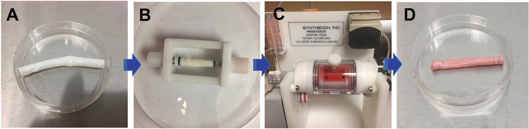
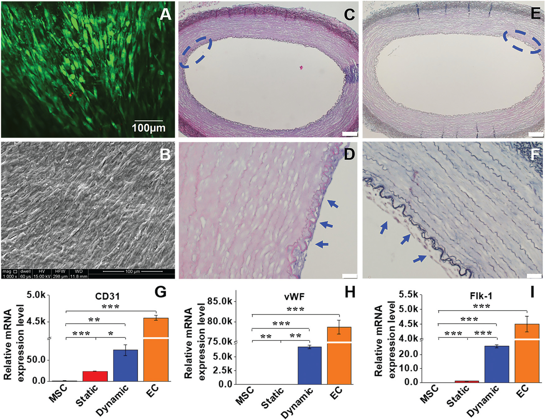
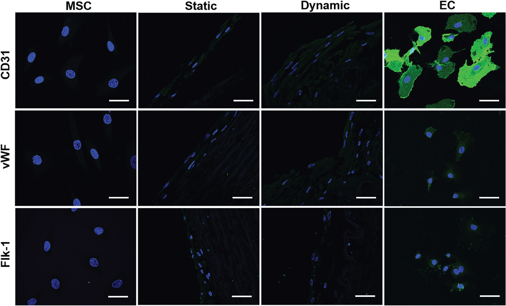
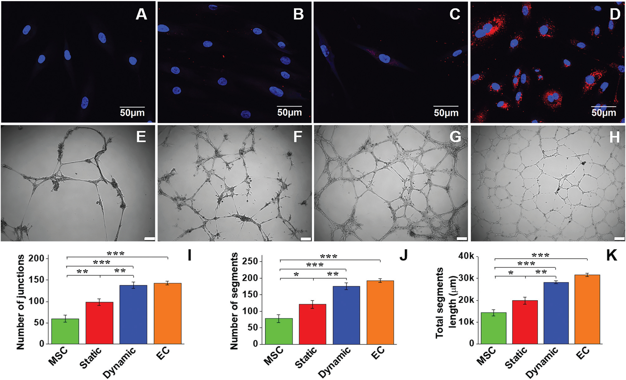
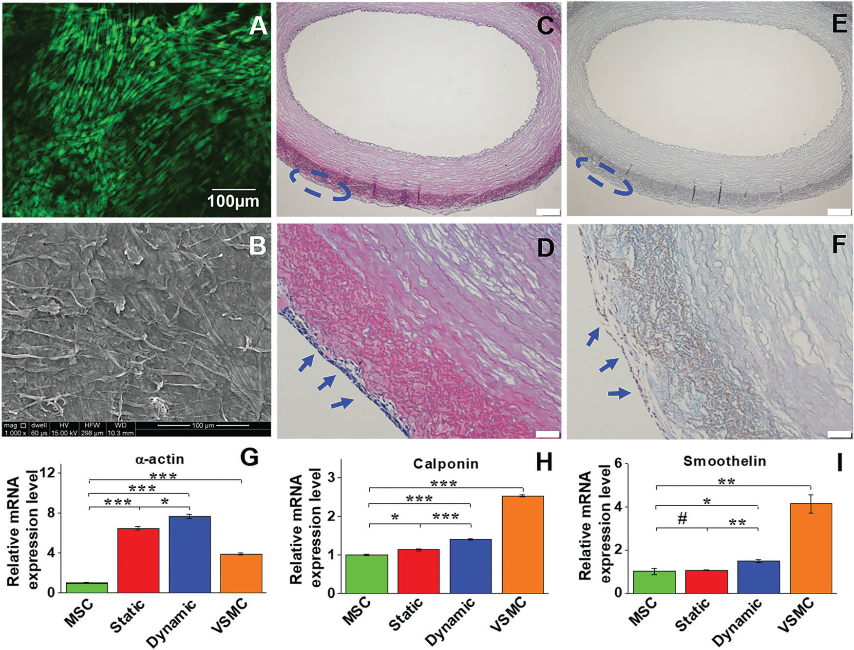
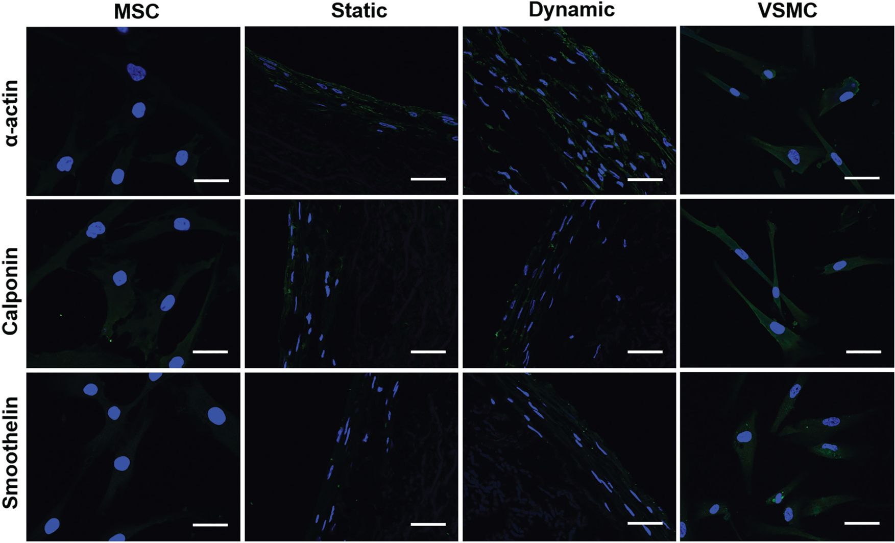
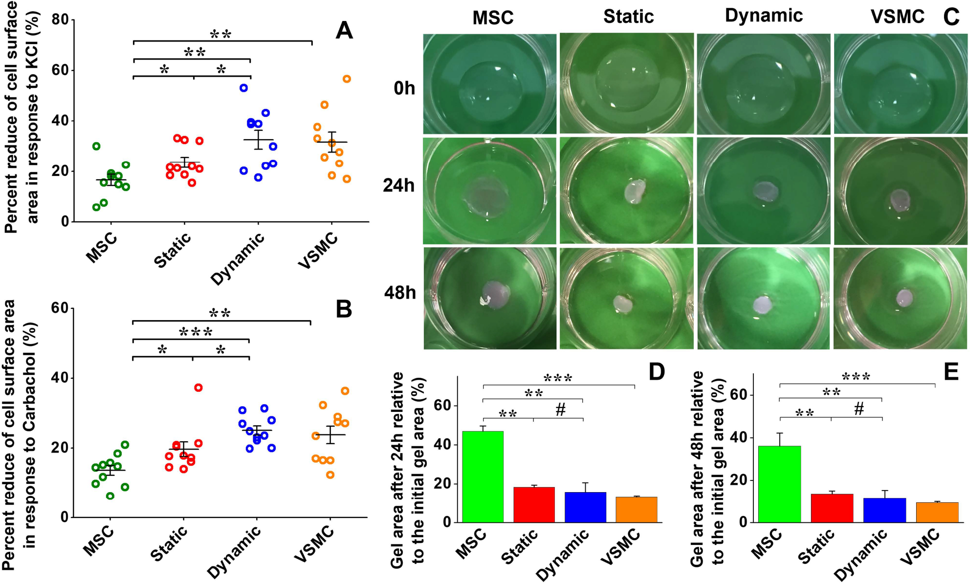
Although a significant number of studies on vascular tissue engineering have been reported, the current availability of vessel substitutes in the clinic remains limited mainly due to the mismatch of their mechanical properties and biological functions with native vessels. In this study, a novel approach to fabricating a vessel graft for vascular tissue engineering was developed by promoting differentiation of human bone marrow mesenchymal stem cells (MSCs) into endothelial cells (ECs) and vascular smooth muscle cells (VSMCs) on a native vascular extracellular matrix (ECM) scaffold in a rotary bioreactor. The expression levels of CD31 and vWF, and the LDL uptake capacity as well as the angiogenesis capability of the EC-like cells in the dynamic culture system were significantly enhanced compared to the static system. In addition, α-actin and smoothelin expression, and contractility of VSMC-like cells harvested from the dynamic model were much higher than those in a static culture system. The combination of on-site differentiation of stem cells towards vascular cells in the natural vessel ECM scaffold and maturation of the resulting vessel construct in a dynamic cell culture environment provides a promising approach to fabricating a clinically applicable vessel graft with similar mechanical properties and physiological functions to those of native blood vessels.


Conflict of interest statement

Conflicts of interest

There are no conflicts to declare.

Figures

Fig. 1
Fig. 1
Schematic of the ex vivo bioreactor incorporated with a decellularized carotid artery scaffold. (A) Decellularized ECM scaffold. (B) MSCs were seeded on either the intimal or adventitial side of the decellularized carotid artery scaffold and mounted on the bioreactor insert. (C) The ECM-MSCs were cultured in a vascular bioreactor with VEGF medium inside the vessel scaffold and TGFβ1 medium outside the vessel scaffold. (D) The tissue engineered blood vessel (TEBV).
Fig. 2
Fig. 2
Cell viability and morphological characteristics, and real time RT-PCR analysis of the EC marker gene expression. (A) MSCs reseeded on the intimal side of decellularized ECM in the dynamic model displayed high cell viability after two weeks. (B) An intact cell layer covered with secreted ECM was observed on the intimal surface of the decellularized scaffolds. (C and D) H&E staining and (E and F) Masson’s trichrome staining confirmed that a dense layer of EC-like cells was formed on the intimal side of the decellularized ECM (blue arrows). Each marker gene was significantly upregulated for both static and dynamic groups compared to the negative control (MSCs), and the transcript expression level of each marker gene in the dynamic culture system was significantly higher than that in the static culture system (G–I). Data are expressed as mean ± SEM (n = 3). *P < 0.05, **P < 0.01, ***P < 0.001.
Fig. 3
Fig. 3
Immunofluorescent staining for EC-specific proteins CD31, vWF, and flk-1. Each marker protein expression level in both the static and dynamic groups was much higher than that in the MSC control group. Both CD31 and vWF expression levels in the dynamic group were stronger than those in the static group. The images represent at least three independent experiments. Scale bars = 50 μm.
Fig. 4
Fig. 4
LDL-uptake assay and Matrigel angiogenesis evaluation of MSC-derived EC-like cells. Negative control MSCs were unable to uptake Dil Ac-LDL (A), whereas positive control ECs exhibited a high capability for Dil Ac-LDL uptake (D). Compared to the static culture group (B), the dynamic culture group showed a much higher Dil Ac-LDL uptake capability (C). Negative control MSCs showed a small amount of capillary network formed on the Matrigel after 24 h (E). In contrast, a dense capillary network was generated by positive control ECs on the Matrigel after 24 h (H). The cells of the static group (F) apparently formed more networks than the MSC group, but much less than the dynamic group (G). Scale bars = 100 μm. Quantitative analysis of the parameters of the capillary network showed that significant differences existed among the four different cell groups (I–K). Data are expressed as mean ± SEM of five randomly selected images (48 cm × 36 cm). *P < 0.05, **P < 0.01, ***P < 0.001.
Fig. 5
Fig. 5
Cell viability and morphology, and mRNA expression of VSMC marker proteins in different experimental groups. Live-dead staining showed that cells cultured on the adventitial side of the vessel scaffold exhibited high viability on day 14 (A). SEM imaging also demonstrated that the cells cultured in the dynamic system were able to form an intact cell layer with uniform topology after two weeks (B). H&E staining (C and D) and Masson’s trichrome staining (E and F) showed multiple layers of VSMC-like cells grown on the adventitial side of the vessel scaffolds (blue arrows). Real time RT-PCR revealed that gene expression of α-actin and calponin was significantly upregulated in each differentiation group compared to the control MSCs (G). The mRNA expression level of each marker protein in the dynamic group was significantly higher than that in the static group, and α-actin expression levels in both the dynamic and static groups were even higher than that in the positive control VSMC (H). Data are expressed as mean ± SEM (n = 3). #P > 0.05, *P < 0.05, **P < 0.01, ***P < 0.001.
Fig. 6
Fig. 6
Immunostaining of VSMC marker proteins for different cell groups. The fluorescence intensity of α-actin in the dynamic group was much higher than that in the static group, and smoothelin in the dynamic group was slightly upregulated compared to the static group. The images represent at least three independent experiments. Scale bars = 50 μm.
Fig. 7
Fig. 7
Functional analysis of MSC-derived VSMC-like cells. Cell area reduction assay showed that MSCs have the lowest contractile capability upon exposure to KCl (A) and carbachol (B), respectively. Both KCl and carbachol induced significant decreases in cell area for the static and dynamic groups compared to the negative control MSCs, and the cell area for the dynamic group was reduced more than that for the static group (A and B). Collagen lattice contraction assay showed that four groups of cells embedded in collagen gel could induce apparent contraction of the collagen gel, and the positive VSMC group exhibited the highest contraction capability among them (C). Quantitative analysis demonstrated that there was no significant difference between the collagen gel areas for the static and dynamic groups (D and E). Data are expressed as mean ± SEM (n = 3). #P > 0.05, *P < 0.05, **P < 0.01, ***P < 0.001.

Similar articles

Cited by

References

    1. Unal B, Critchley JA and Capewell S, J. Epidemiol. Community Health, 2003, 57, 530–535. - PMC - PubMed
    1. Laslett LJ, Alagona P, Clark BA, Drozda JP, Saldivar F, Wilson SR, Poe C and Hart M, J. Am. Coll. Cardiol, 2012, 60, S1–S49. - PubMed
    1. Isenberg BC, Williams C and Tranquillo RT, Circ. Res, 2006, 98, 25–35. - PubMed
    1. Greco Song HH, Rumma RT, Ozaki CK, Edelman ER and Chen CS, Cell Stem Cell, 2018, 22, 608. - PMC - PubMed
    1. Kurobe H, Maxfield MW, Breuer CK and Shinoka T, Stem Cells Transl. Med., 2012, 1, 566–571. - PMC - PubMed

Publication types

MeSH terms

LinkOut - more resources