

doi: 10.1021/jasms.0c00085.
Epub 2020 Apr 21.
Fast Protein Footprinting by X-ray Mediated Radical Trifluoromethylation
Affiliations
- PMID: 32255631
- PMCID: PMC7486011
- DOI: 10.1021/jasms.0c00085
Item in Clipboard
Fast Protein Footprinting by X-ray Mediated Radical Trifluoromethylation
J Am Soc Mass Spectrom.
.
Abstract
Synchrotron radiolysis generates hydroxyl radicals (•OH) that are successful footprinting reagents. Here, we describe a new reagent for the synchrotron platform, the trifluoromethyl radical (•CF3). The radical is produced by •OH displacement of •CF3 from sodium triflinate (Langlois reagent). Upon X-ray beam exposure, the reagent labels proteins extensively without any additional chemicals on a millisecond or shorter time scale. The •CF3 is comparably reactive to •OH and produces footprinting information that complements that of •OH alone. This reagent in combination with •OH should enable novel chemistry for protein footprinting on the synchrotron platform.
Figures
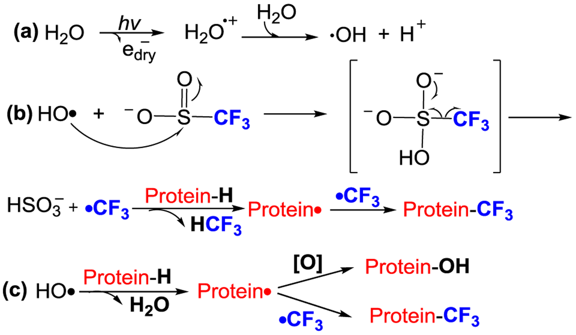
Proposed pathway for X-ray mediated radical trifluoromethylation for protein footprinting.
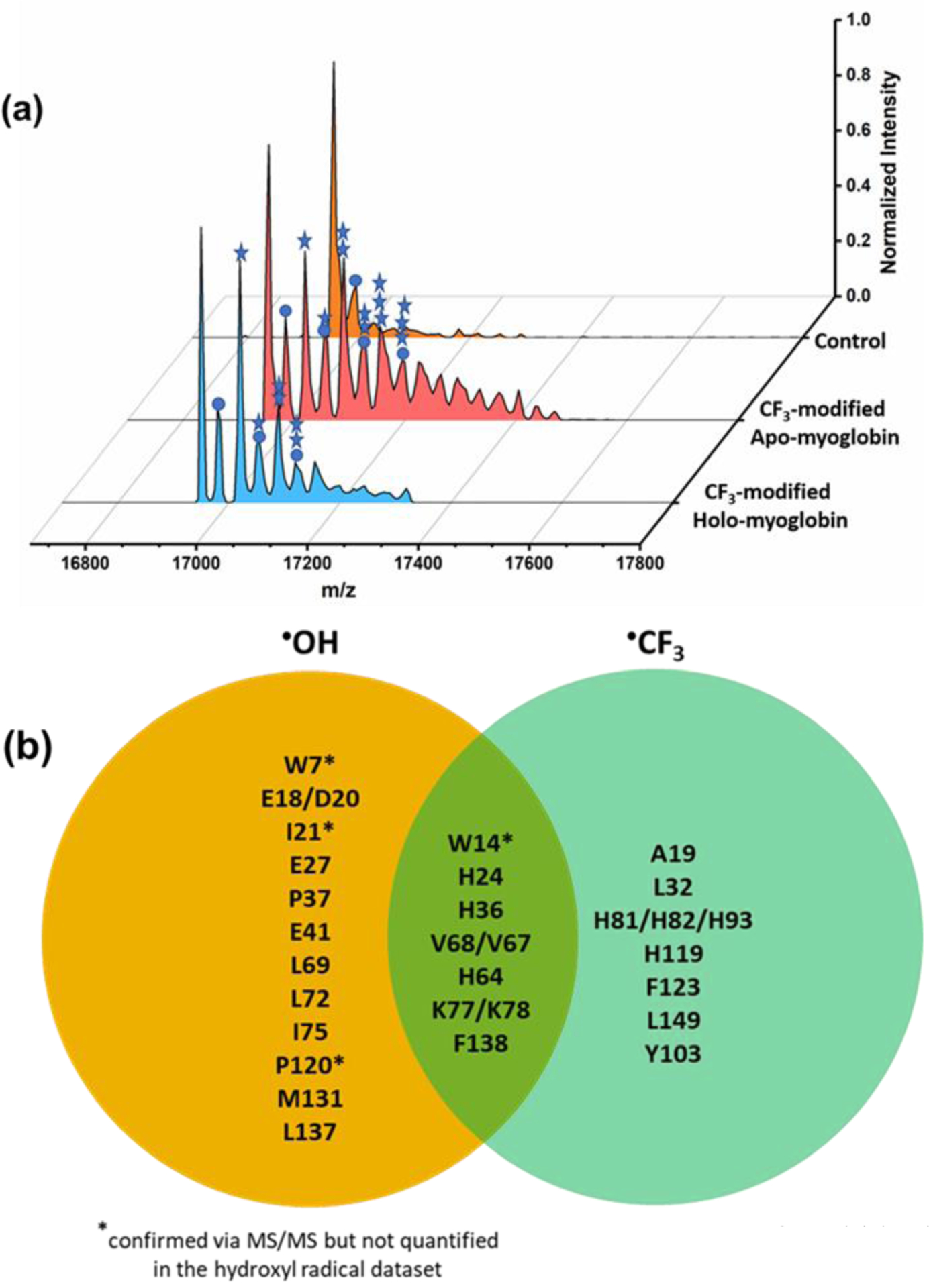
(a) Deconvoluted mass spectra. CF3-modified holo-Mb (front) and apo-Mb (middle) at 25 ms of X-ray irradiation. Apo-Mb (back) without x-ray irradiation shows no CF3-modifications. (b) Stars represent number of trifluoromethylations (+68 Da), and circles represent H substitution by •OH. This figure adapted from Figure S1. Comparison of chemical reactivities for •OH and •CF3 provided by a Venn diagram comparing residues reactive with •OH (in orange) and with •CF3 (in light green); residues in the overlap region are reactive with both •OH and •CF3.
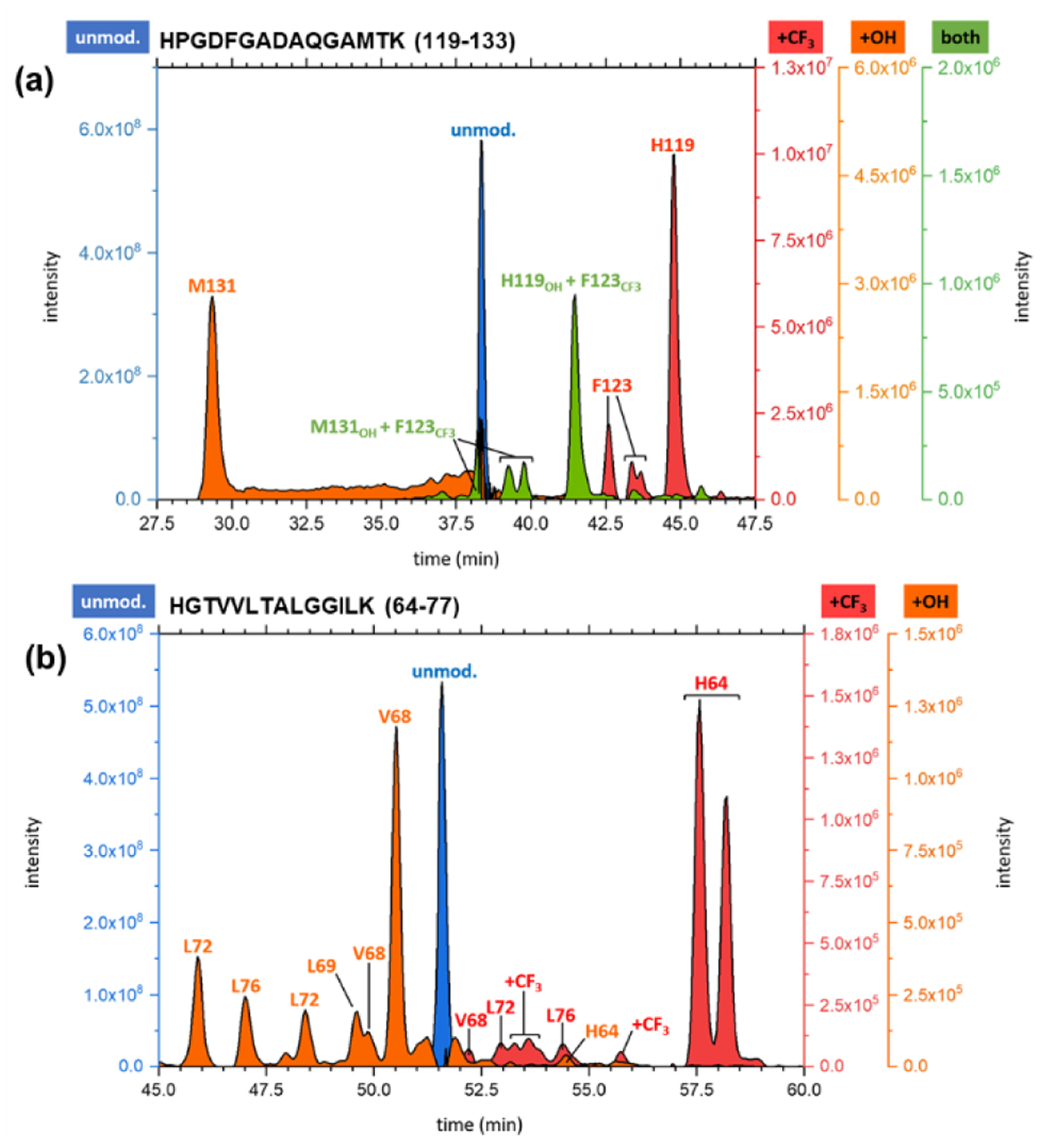
(a) EIC for peptide 119–133 from a 75 ms exposure of holo-Mb for CF3 footprinting on the synchrotron platform. (b) EIC for peptide 64–77 taken from the 50 ms exposure of holo-Mb. EICs for the unmodified (blue), hydroxyl-radical modified (orange), CF3-radical labeled (red), and di-modified (green) peptides confirmed via MS/MS are shown.
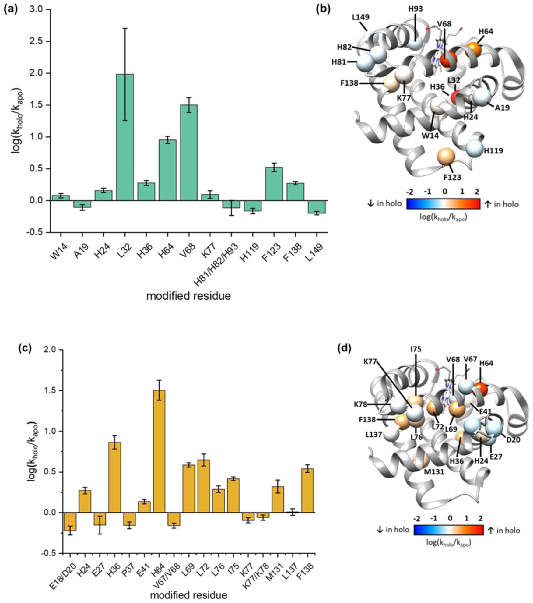
Comparison of X-ray-induced •CF3 and •OH at the AA residue level. Changes in the modification rates respond to heme binding, for the rates of •CF3 modification (a) and of •OH modification (c). Changes mapped on to the structure of holo-Mb (PDB: 1WLA) to identify regions with increased (red), decreased (blue), or unchanged (white) rates of modification upon heme binding, for •CF3 modification (b) and •OH modification (d).
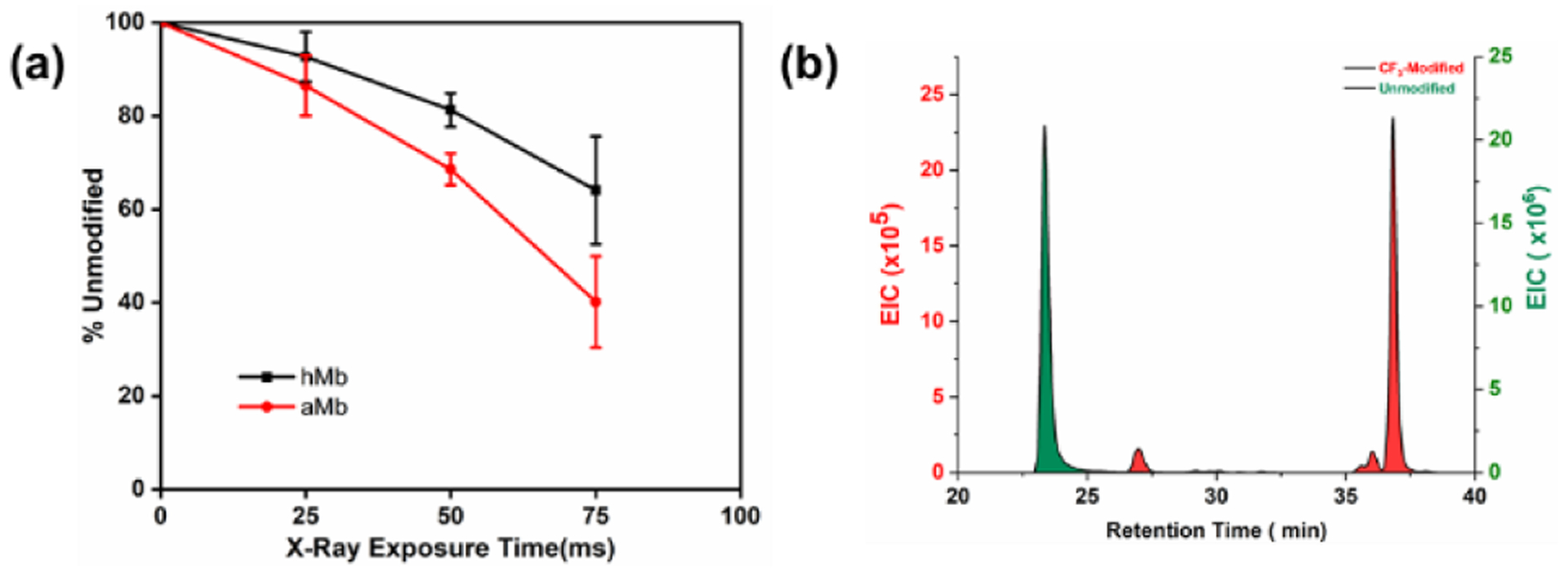
(a) The fraction of unmodified peptide versus exposure time at peptide 80–96. (b)The EIC of peptide 80–96.
Similar articles
-
Multiplex Trifluoromethyl and Hydroxyl Radical Chemistry Enables High-Resolution Protein Footprinting.Anal Chem. 2025 Jan 14;97(1):482-491. doi: 10.1021/acs.analchem.4c04610. Epub 2024 Dec 25. Anal Chem. 2025. PMID: 39720871 Free PMC article.
-
Multiplex Chemical Labeling of Amino Acids for Protein Footprinting Structure Assessment.Anal Chem. 2022 Jul 12;94(27):9819-9825. doi: 10.1021/acs.analchem.2c01640. Epub 2022 Jun 28. Anal Chem. 2022. PMID: 35763792 Free PMC article.
-
Intact mass spectrometry screening to optimize hydroxyl radical dose for protein footprinting.Biochem Biophys Res Commun. 2023 Sep 3;671:343-349. doi: 10.1016/j.bbrc.2023.06.020. Epub 2023 Jun 7. Biochem Biophys Res Commun. 2023. PMID: 37329657 Free PMC article.
-
Protein Footprinting with Radical Probe Mass Spectrometry- Two Decades of Achievement.Protein Pept Lett. 2019;26(1):4-15. doi: 10.2174/0929866526666181128124241. Protein Pept Lett. 2019. PMID: 30484400 Review.
-
Mass Spectrometry-Based Fast Photochemical Oxidation of Proteins (FPOP) for Higher Order Structure Characterization.Acc Chem Res. 2018 Mar 20;51(3):736-744. doi: 10.1021/acs.accounts.7b00593. Epub 2018 Feb 16. Acc Chem Res. 2018. PMID: 29450991 Free PMC article. Review.
Cited by
-
Mass Spectrometry-Based Protein Footprinting for Protein Structure Characterization.Acc Chem Res. 2025 Jan 21;58(2):165-176. doi: 10.1021/acs.accounts.4c00545. Epub 2025 Jan 5. Acc Chem Res. 2025. PMID: 39757421
-
Validated determination of NRG1 Ig-like domain structure by mass spectrometry coupled with computational modeling.Commun Biol. 2022 May 12;5(1):452. doi: 10.1038/s42003-022-03411-y. Commun Biol. 2022. PMID: 35551273 Free PMC article.
-
Workflow for Validating Specific Amino Acid Footprinting Reagents for Protein Higher Order Structure Elucidation.Anal Chem. 2023 Jul 4;95(26):10119-10126. doi: 10.1021/acs.analchem.3c01919. Epub 2023 Jun 23. Anal Chem. 2023. PMID: 37351860 Free PMC article.
-
Structural Investigation of Therapeutic Antibodies Using Hydroxyl Radical Protein Footprinting Methods.Antibodies (Basel). 2022 Nov 14;11(4):71. doi: 10.3390/antib11040071. Antibodies (Basel). 2022. PMID: 36412837 Free PMC article. Review.
-
Protein Structure Prediction with Mass Spectrometry Data.Annu Rev Phys Chem. 2022 Apr 20;73:1-19. doi: 10.1146/annurev-physchem-082720-123928. Epub 2021 Nov 1. Annu Rev Phys Chem. 2022. PMID: 34724394 Free PMC article. Review.
References
-
- Rinas A; Mali VS; Espino JA; Jones LM, Development of a Microflow System for In-Cell Footprinting Coupled with Mass Spectrometry. Anal. Chem 2016, 88, 10052–10058. - PubMed
-
- Maleknia SD; Brenowitz M; Chance MR, Millisecond Radiolytic Modification of Peptides by Synchrotron X-rays Identified by Mass Spectrometry. Anal. Chem 1999, 71, 3965. - PubMed
-
- Kiselar JG; Maleknia SD; Sullivan M; Downard KM; Chance MR, Hydroxyl radical probe of protein surfaces using synchrotron X-ray radiolysis and mass spectrometry. Int. J. Radiat. Biol 2002, 78, 101. - PubMed
Grants and funding
LinkOut - more resources
Full Text Sources
Other Literature Sources
