Review
. 2020 Mar 27:21:183-191.
doi: 10.1016/j.jor.2020.03.043. eCollection 2020 Sep-Oct.

Limb salvage reconstruction: Radiologic features of common reconstructive techniques and their complications

Affiliations
Review

Limb salvage reconstruction: Radiologic features of common reconstructive techniques and their complications

Jose R Perez et al. J Orthop. .

Abstract

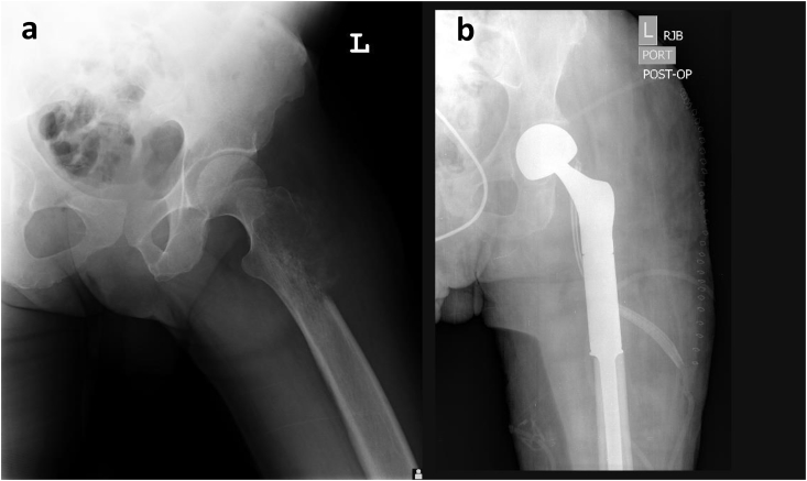
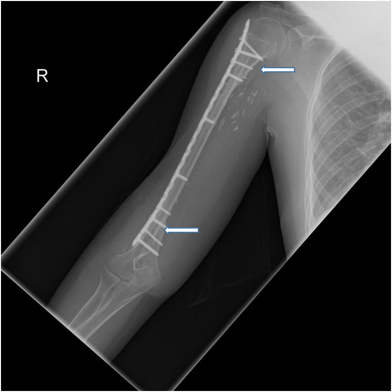
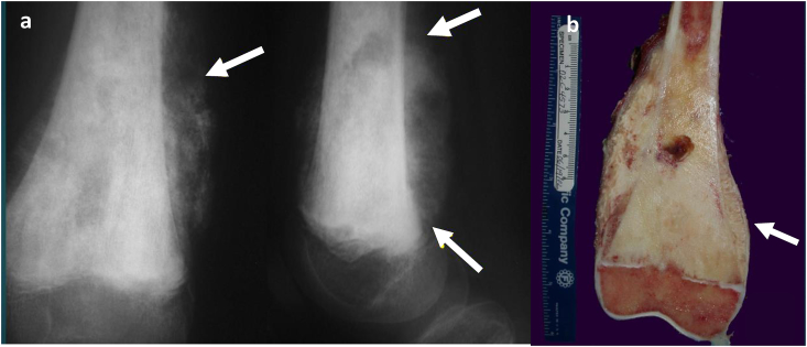
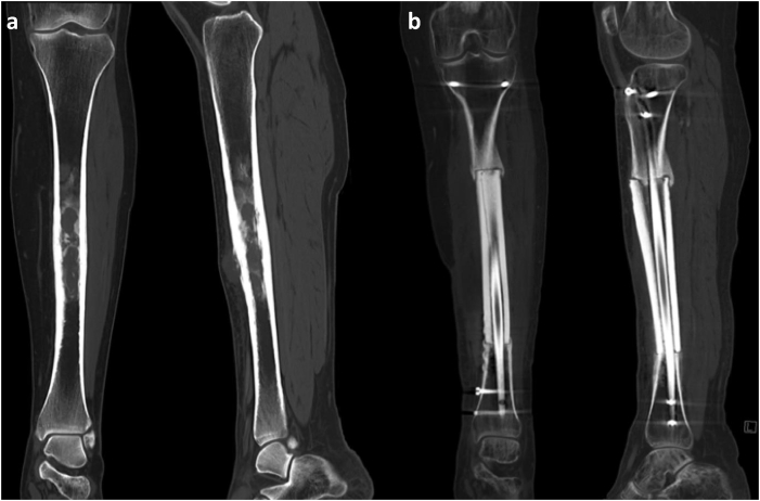
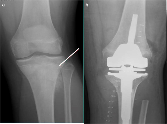
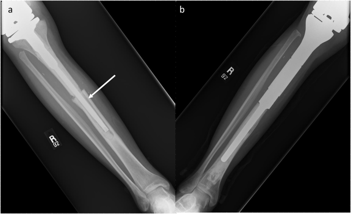
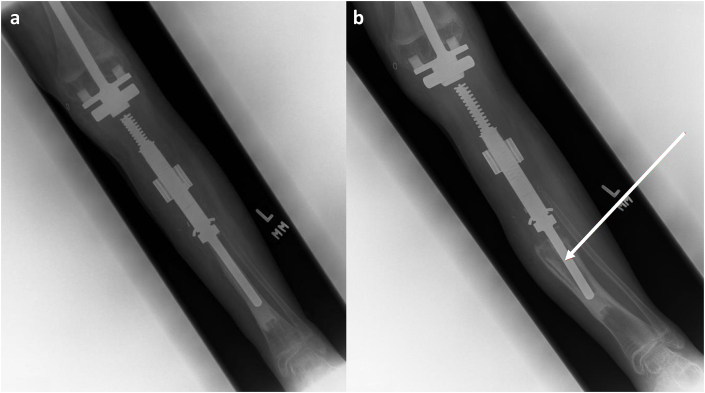
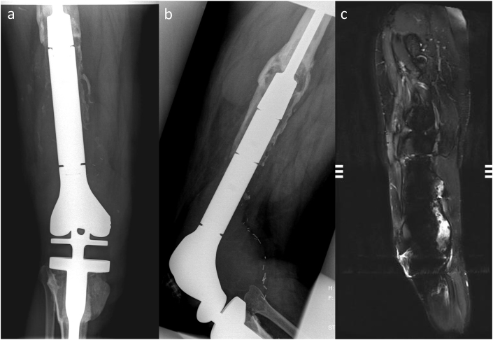
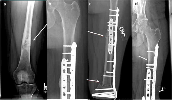
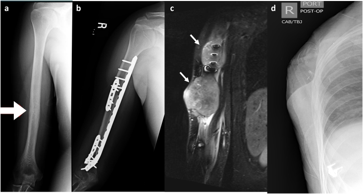
Limb salvage surgery refers to orthopaedic procedures designed to resect tumors and reconstruct limbs. Improvements in managing malignant bone lesions have led to a dramatic shift in limb salvage procedures. Orthopaedic surgeons now employ four main reconstructive procedures: endoprosthesis, autograft, bulk allograft, and allograft prosthetic composite. While each approach has its advantages, each technique is associated with complications. Furthermore, knowledge of procedure specific imaging findings can lead to earlier complication diagnosis and improved clinical outcomes. The aim of this article is to review leading reconstructive options available for limb salvage surgery and present a case series illustrating the associated complications.

Keywords: Allograft; Autograft; Endoprosthesis; Limb salvage.


Conflict of interest statement

All authors declare that they have no conflict of interest. This article does not contain any studies with human participants or animals performed by any of the authors.

Figures

Fig. 1
Fig. 1
(a and b) Proximal Femoral Endoprosthesis. A patient with pathology-confirmed osteosarcoma. (a) Frontal radiographic view of the left pelvis demonstrates lytic lesion (white arrow) involving lateral aspect of the proximal left femur with associated aggressive periosteal reaction. (b) Post-operative radiograph reveals intact femoral endoprosthesis in proper alignment with preservation of native acetabulum.
Fig. 2
Fig. 2
Fibular Allograft Intercalary Reconstruction. A 15-year-old male with history of right proximal humerus osteosarcoma. Frontal post-operative radiographic views of the right humerus demonstrate intact intercalary fibular autograft following resection of diaphyseal osteosarcoma. Note complete incorporation of the graft within native bone at the distal and proximal junctional anastomosis (arrows).
Fig. 3
Fig. 3
(a and b) Distal Femur Bulk Allograft. A patient with osteoblastic osteosarcoma. (a) Frontal and lateral radiographs of the knee demonstrate large mixed sclerotic and lytic lesion with aggressive periosteal reaction (white arrows) with involvement of the distal femoral physis. (b) Gross depiction of the distal femur reveals extensive subperiosteal and physeal involvement (white arrow), necessitating osteoarticular allograft.
Fig. 4
Fig. 4
(a and b) Tibial Diaphyseal Intercalated Allograft Reconstruction. A patient with Ewing sarcoma in the left tibial diaphysis. (a) Coronal and sagittal CT images demonstrate mildly expansile, mixed lytic and sclerotic, marrow-replacing lesion with associated circumferential periosteal and cortical thickening, as well as associated anterior soft tissue component. (b) Coronal and sagittal CT images demonstrate left tibial diaphyseal resection, intercalary graft reconstruction, and myocutaneous flap.
Fig. 5
Fig. 5
(a and b) Tibial Metaphysis Allograft Prosthetic Composite. A patient with osteosarcoma. (a) Pre-operative frontal view demonstrates an aggressive sclerotic lesion in the proximal tibial metaphysis with extension into the physis (white arrow). There is periosteal reaction with fibular extension. (b) Post-operative frontal view shows proximal tibia and fibular head resection with allograft prosthetic composite reconstruction.
Fig. 6
Fig. 6
(a and b) Non-Union of the Graft-Host Junction. A 28-year-old male with history of Ewing sarcoma of the proximal tibia. (a) Frontal radiographic views of the right tibia following APC reconstruction demonstrate non-union of the graft-host junction with lucency around the hardware (white arrow) reflecting osteolysis and hardware loosening. (b) Frontal radiographic view of the right tibia after revision with proximal tibia endoprosthesis.
Fig. 7
Fig. 7
(a and b) Tibial Shaft Aseptic Loosening. A patient with proximal tibia osteosarcoma. (a) Post-operative frontal radiograph demonstrates expandable femorotibial endoprosthesis, allowing limb-lengthening with growth. Follow-up (b) frontal radiograph reveals significant lucency at the bone-cement and metal-cement interfaces with marked posterior and varus angulation of the prosthesis and reactive cortical thickening indicative of aseptic loosening (white arrow).
Fig. 8
Fig. 8
(a, b and c) Femur Periprosthetic Infection. (a) Frontal and (b) lateral radiographs of the right distal femur and proximal tibia with osteolysis surrounding the femoral endoprosthesis and a large, complex effusion at the knee (white arrows). (c) Sagittal STIR MR image demonstrates osteolysis around femoral endoprosthesis with hardware loosening and periprosthetic large enhancing complex collection that extends intra-articularly into the right knee joint pseudocapsule.
Fig. 9
Fig. 9
(a, b, c and d) Proximal and Distal Femoral Shaft Non-Union. A 20-year-old female with pathology-confirmed osteosarcoma. (a) Frontal radiographs of left femur demonstrate mixed lytic and sclerotic aggressive appearing lesion. The lesion spared the femoral epiphysis, allowing resection into the metaphysis to ensure negative margins and (b) use of allograft reconstruction. Follow-up (c) frontal radiograph demonstrates non-union at the proximal and distal graft-junctions (white arrows). Iliac crest bone graft was applied to accelerate healing of the allograft host junction with subsequent healing on (d) 3-month follow-up frontal film (white arrow).
Fig. 10
Fig. 10
(a, b, c and d) Ewing's Sarcoma Recurrence. A 23-year-old male with Ewing sarcoma. (a) Frontal radiograph demonstrates permeative lytic lesions (white arrows) within humeral shaft with surrounding aggressive periosteal reaction and associated soft tissue mass. (b) Frontal radiographs following tumor resection with intercalary allograft placement and no residual tumor. 2-month follow-up (c) Coronal T2 MRI sequences reveal large T2 hyperintense soft tissue masses (white arrows) at the surgical site, encasing the native bone and allograft, consistent with tumor recurrence. The patient refused treatment at this time and returned to his native country. At 7-month follow-up, limb-sparing surgery was no longer feasible due to axillary extension and metastatic spread. (d) Forequarter amputation of the extremity was subsequently performed.

Similar articles

Cited by

References

    1. Dhammi I.K., Kumar S. Osteosarcoma: a journey from amputation to limb salvage. Indian J Orthop. 2014;48(3):233–234. - PMC - PubMed
    1. DiCaprio M.R., Friedlaender G.E. Malignant bone tumors: limb sparing versus amputation. J Am Acad Orthop Surg. 2003;11(1):25–37. - PubMed
    1. Yang Y., Han L., He Z. Advances in limb salvage treatment of osteosarcoma. J Bone Oncol. 2018;10:36–40. - PMC - PubMed
    1. Chauhan A., Joshi G.R., Chopra B.K., Ganguly M., Reddy G.R. Limb salvage surgery in bone tumors: a retrospective study of 50 cases in a single center. Indian J Surg Oncol. 2013;4(3):248–254. - PMC - PubMed
    1. Solooki S., Mostafavizadeh Ardestani S.M., Mahdaviazad H., Kardeh B. Function and quality of life among primary osteosarcoma survivors in Iran: amputation versus limb salvage. Musculoskelet Surg. 2017 - PubMed

LinkOut - more resources