Review
. 2020 Mar 19:13:785-810.
doi: 10.2147/DMSO.S234992. eCollection 2020.

The Exercise Training Modulatory Effects on the Obesity-Induced Immunometabolic Dysfunctions

Affiliations
Review

The Exercise Training Modulatory Effects on the Obesity-Induced Immunometabolic Dysfunctions

Nakisa Soltani et al. Diabetes Metab Syndr Obes. .

Abstract

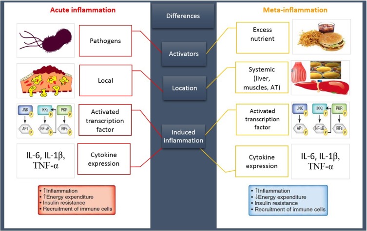
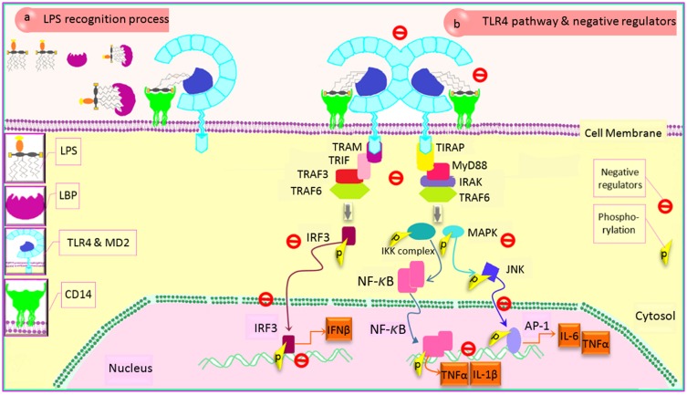
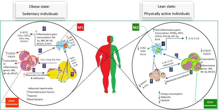
Reduced physical activity rate in people's lifestyle is a global concern associated with the prevalence of health disorders such as obesity and metabolic disturbance. Ample evidence has indicated a critical role of the immune system in the aggravation of obesity. The type, duration, and production of adipose tissue-released mediators may change subsequent inactive lifestyle-induced obesity, leading to the chronic systematic inflammation and monocyte/macrophage (MON/MФ) phenotype polarization. Preliminary adipose tissue expansion can be inhibited by changing the lifestyle. In this context, exercise training is widely recommended due to a definite improvement of energy balance and the potential impacts on the inflammatory signaling cascades. How exercise training affects the immune system has not yet been fully elucidated, because its anti-inflammatory, pro-inflammatory, or even immunosuppressive impacts have been indicated in the literature. A thorough understanding of the mechanisms triggered by exercise can suggest a new approach to combat meta-inflammation-induced metabolic diseases. In this review, we summarized the obesity-induced inflammatory pathways, the roles of MON/MФ polarization in adipose tissue and systemic inflammation, and the underlying inflammatory mechanisms triggered by exercise during obesity.

Keywords: adipose tissue; exercise training; immune system; macrophage polarization; meta-inflammation; obesity; toll-like receptors.


Conflict of interest statement

The authors have no conflicts of interest to declare.

Figures

Figure 1
Figure 1
A classic inflammation VS meta-inflammation. Both pathways induce an inflammatory state following the upregulation of same inflammatory genes and cytokines. An acute inflammation has been promoted by invasive exogenous pathogens leading to the stimulation of immune system that induces a high-intensity short-term local inflammation. In contrast, a meta-inflammation has been developed by the excess nutrient, endogen stimulants, causing immune system provocation and promoting a chronic LGSI. Abbreviations: AT, adipose tissue; IL, interleukin; TNFα, tumor necrosis factor α; JNK, c-Jun N-terminal kinase; IKK, inhibitor of kappa light polypeptide gene enhancer in B-cells kinase; PKR, protein kinase R; NF-κB, nuclear factor kappa-light-chain-enhancer of activated B cells; IRF, interferon regulatory factor.
Figure 2
Figure 2
LPS transferring pathway and modulation of TLR4 signaling cascade. (a) TLR4-MD2 complex and LPS recognizing: TLR4 signaling cascade is activated following LPS sensing mediated by three different protein activities, causing both ligand recognizing and transferring to receptor. The LBP-LPS complex is recognized by CD14, TLR4 co-receptor, leading to TLR4 cascade activation. (b) TLR4 protein adaptors and negative regulators: TLR4 signaling necessitates recruiting two main adaptors, including MyD88 and TRIF. Activation of other mediators causes the phosphorylation of some proteins which leads to the translocation of inflammatory transcription factors into nucleus, inducing effective inflammatory genes upregulation. Modulating of TLR4 is mediated by the negative regulator proteins in different stages (TLR4 sensing ligand, CD14, adaptor recruitment, transcriptional factor activation, transcriptional factor translocation, gene transcription) to either control the excessive response of TLR4 or suppress its activities at an effective time following an inflammatory response. Abbreviations: TLR4, toll-like receptor 4; LPS, lipopolysaccharide; LBP, LPS-binding protein; CD, cluster of differentiation; MD2, myeloid differentiation factor 2; MyD88, myeloid differentiation factor 88; TRIF, TIR domain-containing adaptor-inducing IFN-β; TIRAP, TIR domain-containing adaptor protein; TRAM, TRIF-related adaptor molecule; IRAK, IL-1R-associated kinase; TRAF6, TNF-receptor-associated factor 6; IKK, inhibitor of kappa light polypeptide gene enhancer in B-cells kinase; IL, interleukin; MAPK, mitogen-activated protein kinase; TNFα, tumor necrosis factor α; JNK, c-Jun N-terminal kinase; AP-1, activator protein 1; NF-κB, nuclear factor kappa-light-chain-enhancer of activated B cells; IRF, interferon regulatory factor; p, phosphorylation.
Figure 3
Figure 3
Physical activity alters circulating monocyte features and obese adipose tissue macrophage distinction. Obese state: The excess nutrients-caused adipocyte hypertrophy and cell death induce the micro hypoxia in AT. ① Although MФs accumulation is increased, constantly elevated levels of circulating non-esterified fatty acid reveal an inefficient function of MФs to maintain the AT homeostasis. These complex interactions of the immune system and adipocyte cause the release of inflammatory cytokines into the blood. Both circulating cytokines and the fatty-acids provide a chemoattractive environment. ② The increased CCL2 release and hypoxia result ③ the MON recruitment and more intensive infiltration into the AT. In AT, this inflammatory condition promotes M1 polarization leading to ④ higher number of surface receptors such as TLR4 and CCR2 which activate the influential inflammatory transcription factors, resulting in the upregulation of several inflammatory cytokine genes such as CCL and CXCL family, IFNγ, IL-6/12, and TNFα. ⑤ Circulating MONs are affected by releasing the abundant inflammatory cytokines and FFAs into the bloodstream. They might be altered toward the inflammatory types due to the increase of inflammatory receptors and, consequently, activation of inflammatory signaling cascades, causing the upregulation of more inflammatory genes. This impaired feed-forward cycle aggravates the inflammatory status in individuals with obesity. Lean state: Exercise training improves the inflammatory state through the inhibition of AT expansion (increasing energy consumption and modulation of inflammatory cascade activities (anti-inflammatory cytokine expression). It seems that the improvement of the inflammatory condition in either AT or bloodstream is induced by the reduction of the chemoattractant factors like CCL2 and FFAs. This has influenced the MФ polarization pathway toward M2. Consequently, it leads to a decrease in both the adipocyte death and the expression of inflammatory cytokine receptors on the surface of cell membrane, which resulted in the downregulation of inflammatory pathways. The circulating MONs which experience the anti-inflammatory condition obtain the features of anti-inflammatory MON subsets. Moreover, producing the strong anti-inflammatory myokines could interrupt these pathways following the exercise training, resulting in the improvement of obesity induced-inflammatory cycle. Abbreviations: TLR4, toll-like receptor 4; CD, cluster of differentiation; fet-A, fetuin-A; CCR2, C-C chemokine receptor type 2; HIF1α, hypoxia-inducible factor 1 α; JNK, c-Jun N-terminal kinase; NF-κB, nuclear factor kappa-light-chain-enhancer of activated B cells; IRF, interferon regulatory factor; STAT, signal transducer and activator of transcription; IL, interleukin; CCL, C-C motif chemokine ligand; CXCL, C-X-C motif chemokine ligand, TNFα, tumor necrosis factor α; IFNγ, interferon regulatory factor γ; SOCS, suppressor of cytokine signaling; CCR, C-C motif chemokine receptor; FFA, free fatty-acid; SFA, saturated fatty-acid; PGC1β, peroxisome proliferator-activated receptor co-activator 1 β; PPARγ, peroxisome proliferator-activated receptor γ; KLF4, kruppel like factor 4.

Similar articles

Cited by

References

    1. Man K, Kutyavin VI, Chawla A. Tissue immunometabolism: development, physiology, and pathobiology. Cell Metab. 2017;25(1):11–26. doi:10.1016/j.cmet.2016.08.016 - DOI - PMC - PubMed
    1. Hotamisligil GS, Shargill NS, Spiegelman BM. Adipose expression of tumor necrosis factor-: direct role in obesity-linked insulin resistance. Sci NY Washinton. 1993;259:87. doi:10.1126/science.7678183 - DOI - PubMed
    1. Weisberg SP, McCann D, Desai M, Rosenbaum M, Leibel RL, Ferrante AW. Obesity is associated with macrophage accumulation in adipose tissue. J Clin Invest. 2003;112(12):1796–1808. doi:10.1172/JCI200319246 - DOI - PMC - PubMed
    1. Makki K, Froguel P, Wolowczuk I. Adipose tissue in obesity-related inflammation and insulin resistance: cells, cytokines, and chemokines. ISRN Inflammation. 2013;2013:1–12. doi:10.1155/2013/139239 - DOI - PMC - PubMed
    1. Ouchi N, Parker JL, Lugus JJ, Walsh K. Adipokines in inflammation and metabolic disease. Nat Rev Immunol. 2011;11(2):85–97. doi:10.1038/nri2921 - DOI - PMC - PubMed