Review
. 2020 Mar 17:2020:3830212.
doi: 10.1155/2020/3830212. eCollection 2020.

Molecular and Cellular Pathways Contributing to Joint Damage in Rheumatoid Arthritis

Affiliations
Review

Molecular and Cellular Pathways Contributing to Joint Damage in Rheumatoid Arthritis

Qinghua Fang et al. Mediators Inflamm. .

Abstract

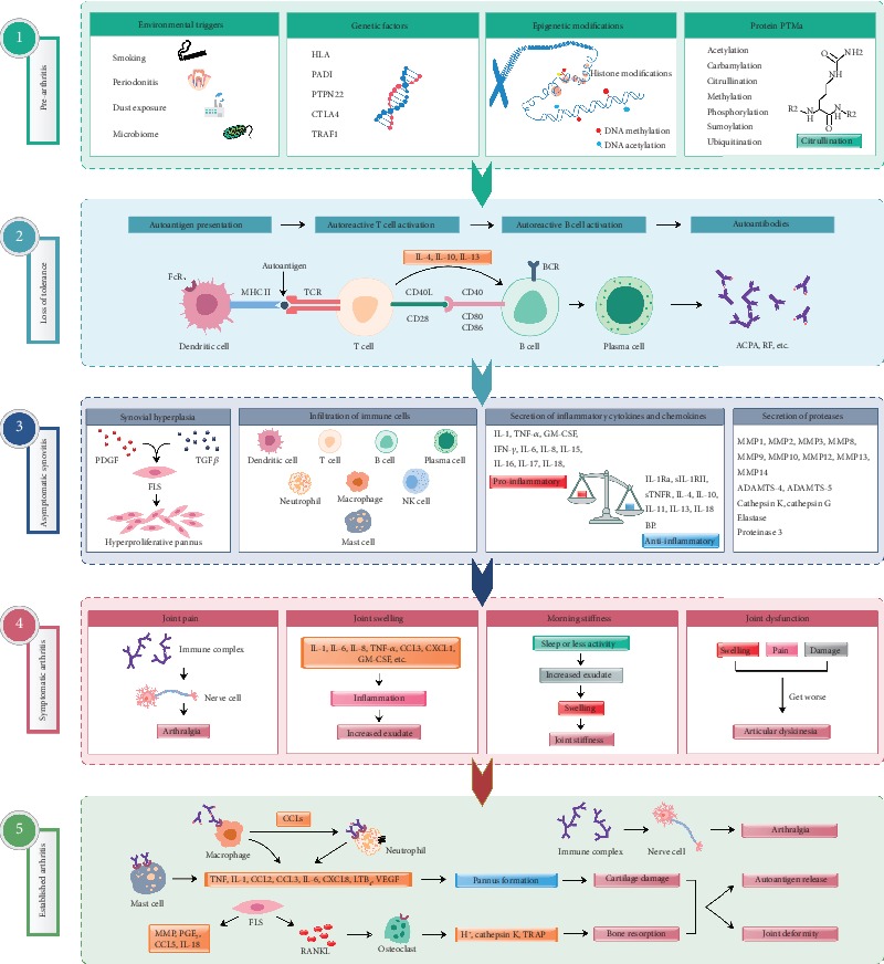
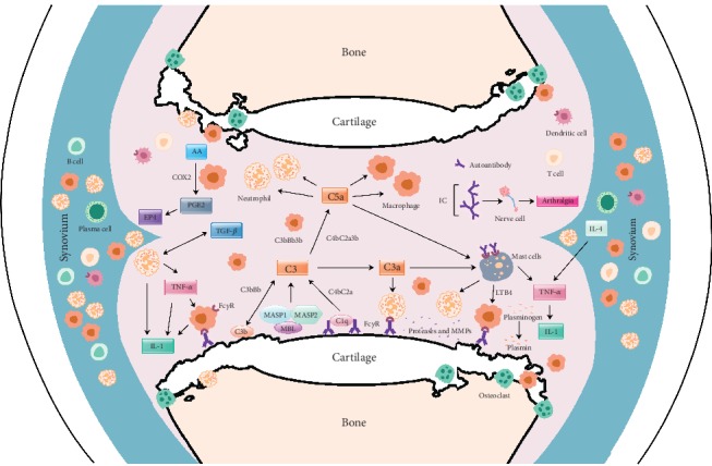
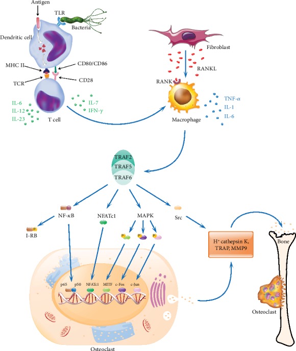
Rheumatoid arthritis is a chronic autoimmune syndrome associated with several genetic, epigenetic, and environmental factors affecting the articular joints contributing to cartilage and bone damage. Although etiology of this disease is not clear, several immune pathways, involving immune (T cells, B cells, dendritic cells, macrophages, and neutrophils) and nonimmune (fibroblasts and chondrocytes) cells, participate in the secretion of many proinflammatory cytokines, chemokines, proteases (MMPs, ADAMTS), and other matrix lysing enzymes that could disturb the immune balance leading to cartilage and bone damage. The presence of autoantibodies preceding the clinical onset of arthritis and the induction of bone erosion early in the disease course clearly suggest that initiation events damaging the cartilage and bone start very early during the autoimmune phase of the arthritis development. During this process, several signaling molecules (RANKL-RANK, NF-κB, MAPK, NFATc1, and Src kinase) are activated in the osteoclasts, cells responsible for bone resorption. Hence, comprehensive knowledge on pathogenesis is a prerequisite for prevention and development of targeted clinical treatment for RA patients that can restore the immune balance improving clinical therapy.


Conflict of interest statement

The authors declare no conflict of interest.

Figures

Figure 1
Figure 1
Different phases in RA pathogenesis. (1) Genetic, epigenetic, and environmental factors contribute to arthritis progression. Multiple environmental risk factors (for example, smoking, pollutants, or microbes), when come in contact with the mucosal sites, are most likely responsible for causing local inflammatory events and immune system activation inducing epigenetic modifications and protein posttranslational modifications (PTMs) [59], before crossing the threshold to trigger disease in genetically vulnerable people. (2) Dendritic cells presenting altered self or related peptides to T cells (breakdown of tolerance mechanisms) leads to the activation of T and B cells effectuating synthesis of cytokines and autoantibodies. Progressively, these autoantibodies are produced more and more, which recognize several neoepitopes by the process of epitope spreading, and gets overt during the onset of the clinical disease [1]. (3) Disease development involves autoimmune responses against both posttranslationally modified and unmodified self-antigens, which starts many years before the subclinical synovitis and appearance of clinical symptoms [60]. (4) Autoantibodies induced during this preclinical phase can also be responsible for bone erosion and pain. Before the onset of inflammation, these alterations could reduce overall functions of the joints. After the autoantibodies start binding to different epitopes and form immune complexes, inflammation in the synovium and development of arthritis ensue. (5) Antibody-induced cartilage and bone changes, if minor, resolve without any considerable damages. However, if left untreated or in the presence of continuous external stimuli, these changes can give rise to chronic inflammation, joint destruction, and disability [59]. Arthritis is associated with both local as well as systemic pathological manifestations. ACPA: anticitrullinated protein antibody; ADAMTS: a disintegrin and metalloproteinase with thrombospondin motifs; BCR: B cell receptor; CCL: c-c motif chemokine ligand; CXCL: C-X-C motif chemokine ligand; CTLA4: cytotoxic T-lymphocyte-associated protein 4; FcR: Fc receptor; FLS: fibroblast-like synoviocytes; GM-CSF: granulocyte-macrophage colony-stimulating factor; HLA: human leukocyte antigen; IFN-γ: interferon gamma; IL: interleukin; IL-1Ra: interleukin-1 receptor antagonist; IL-18BP: interleukin-18-binding protein; LTB4: leukotriene B4; MMP: matrix metalloproteinase; MHC II: major histocompatibility complex class II; NK cell: natural killer cell; PADI: peptidyl arginine deiminase; PDGF: platelet-derived growth factor; PGE2: prostaglandin E2; PTPN22: protein tyrosine phosphatase nonreceptor type 22; RANKL: receptor activator of nuclear factor kappa B ligand; RF: rheumatoid factor; sIL-1RII: soluble interleukin 1 receptor II; sTNFR: soluble tumor necrosis factor receptors; TCR: T cell receptor; TGFβ: transforming growth factor β; TNF-α: tumor necrosis factor α; TRAF1: TNF receptor-associated factor 1; VEGF: vascular endothelial growth factor.
Figure 2
Figure 2
Likely interactions of molecules and factors in the antibody mediated joint inflammation. Upon binding to joint antigens or deposited as immune complexes on the cartilage surface, autoantibodies initiate inflammation-dependent and inflammation-independent activities, which culminate in the direct damage to the cartilage and bone. Activation of complement cascades by autoantibodies leads to the release of anaphylatoxins (C3a, C5a), attracting FcR-bearing immune cells to the inflammation foci, which in turn get more activated and secrete cytokines that can further activate resident nonimmune cells in the joint. All these cells in the inflamed joint secrete more inflammatory mediators and extracellular matrix lysing enzymes that could destroy the cartilage and bone. AA: arachidonic acid; C1q, C2a, C3, C3a, C4b and C5a and B (factor B): complement components; CCL3: chemokine (C-C motif) ligand 3; COX2: cyclooxygenase-2; EP4: prostaglandin receptor; MASP: mannose-associated serine protease; MBL: mannose-binding lectin; IC: immune complex; IL: interleukin; LTB4: leukotriene B4; FcγR: Fc gamma receptors; PGE2: prostaglandin E2; TGF: transforming growth factor; TNF: tumor necrosis factor.
Figure 3
Figure 3
Signaling pathways in osteoclast activation. During RA pathogenesis, antigen-presenting cells after uptake of an autoantigen or pathogenic molecules process and present antigenic determinants on their cell surface in conjunction with arthritis-permissible HLA/MHC class II molecules, which activate differentiation of T cells into different subphenotypes. The activated T cells secrete various cytokines like IL-6, IL-7, IL-10, IL-12, IL-17, IL-23, and IFN-γ. These cytokines modulate macrophages to secrete various pro- and/or anti-inflammatory cytokines and other inflammatory mediators. Upon exposure to the inflammatory cytokines, fibroblast-like synoviocytes express RANKL, which binds with its receptor (RANK) present on the cell surface of activated macrophages initiating the RANK/RANKL pathway through TRAF 2, 5, and 6 proteins, which leads to the activation of downstream NF-κB, MAPK, NFATc1, and Src signaling cascades. These factors after translocation initiate the expression of genes like TRAP, CtsK, and MMP-9 in the nucleus, which promote osteoclastogenesis and bone resorption. TLR: toll-like receptor; TCR: T cell receptor; CtsK: cathepsin K; I-κB: inhibitor of the NF-κB transcription factor; IL: interleukin; IFN-γ: interferon gamma; MAPK: mitogen-activated protein kinase; MHC II: major histocompatibility complex II; MITF: microphthalmia-associated transcription factor; MMP 9: matrix metalloproteinase 9; NFATc1: nuclear factor of activated T cells, calcineurin-dependent 1; NF-κB: nuclear factor kappa B; p50 and p65: REL-associated proteins (also called NF-κB1 and RelA) involved in NF-κB heterodimer formation and nuclear translocation and activation; RANK: receptor activator of nuclear factor-κB; RANKL: receptor activator of nuclear factor κB ligand; Src: intracellular non-receptor tyrosine kinase; TRAF: TNF receptor-associated factor; TRAP: tartrate-resistant acid phosphatase. c-Jun and c-Fos form the early response transcription factor, AP-1.

References

    1. McInnes I. B., Schett G. Pathogenetic insights from the treatment of rheumatoid arthritis. Lancet. 2017;389(10086):2328–2337. doi: 10.1016/S0140-6736(17)31472-1. - DOI - PubMed
    1. Orr C., Vieira-Sousa E., Boyle D. L., et al. Erratum: Synovial tissue research: a state-of-the-art review. Nature Reviews Rheumatology. 2017;13(10):p. 630. doi: 10.1038/nrrheum.2017.161. - DOI - PubMed
    1. Fang Q., Ou J., Nandakumar K. S. Autoantibodies as diagnostic markers and mediator of joint inflammation in arthritis. Mediators of Inflammation. 2019;2019:22. doi: 10.1155/2019/6363086.6363086 - DOI - PMC - PubMed
    1. Harre U., Lang S. C., Pfeifle R., et al. Glycosylation of immunoglobulin G determines osteoclast differentiation and bone loss. Nature Communications. 2015;6(1):p. 6651. doi: 10.1038/ncomms7651. - DOI - PMC - PubMed
    1. Burmester G. R., Feist E., Dorner T. Emerging cell and cytokine targets in rheumatoid arthritis. Nature Reviews Rheumatology. 2014;10(2):77–88. doi: 10.1038/nrrheum.2013.168. - DOI - PubMed