. 2020 Feb 28;22(8):1448-1458.
doi: 10.1039/c9ce01587f. Epub 2020 Feb 5.

In situ flow cell platform for examining calcium oxalate and calcium phosphate crystallization on films of basement membrane extract in the presence of urinary 'inhibitors'

Affiliations

In situ flow cell platform for examining calcium oxalate and calcium phosphate crystallization on films of basement membrane extract in the presence of urinary 'inhibitors'

Cary A Kuliasha et al. CrystEngComm. .

Abstract

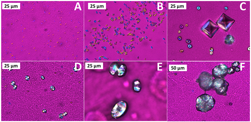
A significant portion of the population suffers from idipoathic calcium oxalate (CaOx) kidney stones, and current clinical treatments of stones have limited lasting success with a high rate of patients suffering from reoccurring stones. Understanding the role of physiologically relevant urinary species on the formation, aggregation, and growth of CaOx crystals can allow for better understanding of this complex biomineralization process and lead to more effective clinical treatments. Our prior work has focused on developing a two-stage model system, where the first stage emulates the formation of Randall's plaque, and the second stage examines the influence of the plaque on overgrowth of CaOx into a stone. Herein, we report on the development of an easy-to-use flow-cell platform that utilizes basement membrane extract (BME) as a biologically relevant crystallization substrate to study the influence of urinary 'inhibitors' on the in situ formation and growth of CaOx on BME under flow conditions. Magnesium, citrate, and osteopontin were studied because of their known ability to inhibit CaOx formation, but their influence also led to interesting modifications to the terminal crystal habit. Magnesium had little to no effect on the CaOx crystallization, but both citrate and osteopontin resulted in significant changes to the crystallization kinetics and the terminal crystal habits. Triply inhibited artificial urine solutions resulted in CaOx monohydrate formations that resembled physiological stones, and the in situ platform allowed for morphogenesis to be dynamically monitored. The BME was also used in a two-stage model system to first grow CaP that mimicked Randall's plaques, whereby the impact of the CaP crystallizing surface on CaOx formation could be studied. It was found that the CaP surface did not result in any significant changes in CaOx crystal formation or growth indicating that the urinary inhibitors and the basement membrane substrate were the dominant factors in modulating CaOx crystallization. It was also found that the basement membrane surface promoted the attachment and/or nucleation and growth of both CaOx and CaP crystals compared to bare glass surfaces, thereby enabling easy study of the urinary inhibitors. The work presented here has elucidated the terminal growth habit of different COM structures and has provided an easy to use platform that can be widely adopted by the kidney stone and other crystallization communities.


Conflict of interest statement

Conflicts of interest There are no conflicts to declare

Figures

Figure 1.
Figure 1.
CaOx mineralization of a collagen sponge with pre-adsorbed CS. (A-B) SEM micrographs of CaOx deposits on the CS-coated collagen sponge in a solution of AU (no soluble polymer additive). (B) Higher magnification of boxed region in (A) shows the spherules are comprised of densely stacked layers of crystals. A few small aggregates of crystals are also present. The asterisk in (A) indicates the region of interest selected for the EDS spectrum in the insert, which shows the stacked crystals are composed of CaOx. The tabular habit suggests these are twinned COM crystals. (C) Another region shows similar stacks of COM tablets as well as some rosette type aggregates, and a few small COD crystals with the typical bipyramid habit. (D) The control reaction, with CS adsorbed onto a glass slide (no collagen), formed a few COD bipyramids, many COM tablets that were either isolated or within rosette aggregates, but the densely layered stacked spherules were not seen.
Figure 2.
Figure 2.
Crystallization flow-cell assembly. (A) Schematic of flow-cell footprint highlighting the placement of the BME, gelation at 37 °C, and respective regions of interest for crystallization. (B) 3D schematic of the flow-cell assembly. (C) Placement of the flow cell below the ultra-long-working-distance objective of a polarized light microscope for in situ analysis of crystal growth at RT.
Figure 3.
Figure 3.
Representative micrographs of Groups 1-2 (A-B), and Group 3 (C-D) using polarized light (with gypsum λ-plate) and SEM, showing relevant CaOx crystal habits. Group 1-2 formed COM dumbbells and coffins and COD bipyramids while group 3 formed COM tablets and COD bipyramids.
Figure 4.
Figure 4.
Raman spectra of COM and COD crystals. All COM and COD crystal habits were distinguished both by Raman and visual inspection.
Figure 5.
Figure 5.
Polarized light micrographs (with gypsum λ-plate) of non-traditional COM structures found in Group 4, showing the onset of crystal formation and growth to COM spherules. (A) crystallization onset as micron-sized droplets; (B-C) growth to COM donuts with distinct dimple structure; (D-E) continued growth and shrinking of central dimple; and (F) fully formed spherule.
Figure 6.
Figure 6.
SEM images of Group 4 non-traditional COM structures. (A-F) Stages of growth that correlate with the relative stages of growth seen in Fig. 5. Insets show EDS spectra of CaOx structures (*). (A) Initial crystalline formation with visible depression on the (100) face that (B-E) starts to slowly fill in with additional crystal growth resulting in (F) fully formed COM spherules.
Figure 7.
Figure 7.
Representative SEM images of fully formed Group 4 COM spherules that closely resemble the morphological characteristics reported for physiological kidney stones.
Figure 8.
Figure 8.
Summary of CaOx crystal habits formed by the four AU groups and their respective crystal planes/directions of interest. (A) COM dumbbell, (B) COM coffin, (C) COD bipyramid, (D) COM tablet, (E) COM spherule (F) COM donut, and (G) extended COD bipyramid.
Figure 9.
Figure 9.
CaOx formation on CaP mineralized BME film using (A-B) Group 1 and (C-D) Group 4 solutions. (A-B) Group 1 resulted in COM dumbbells and coffins and COD bipyramids. (C-D) Group 4 resulted in COM donuts/spherules and tablets and COD bipyramids. CaP did not significantly impact the crystals habits formed relative to the BME flow experiments discussed previously. SEM insets show EDS of (B) CaP and (D) CaOx regions (*)

Similar articles

Cited by

References

    1. Parmar MS, BMJ, 2004, 328, 1420–1424. - PMC - PubMed
    1. Coe FL, Evan A and Worcester E, J. Clin. Invest, 2005, 115, 2598–2608. - PMC - PubMed
    1. Pearle MS, Goldfarb DS, Assimos DG, Curhan G, Denu-Ciocca CJ, Matlaga BR, Monga M, Penniston KL, Preminger GM, Turk TMT and White JR, J. Urol, 2014, 192, 316–324. - PubMed
    1. Daudon M, Bazin D and Letavernier E, Urolithiasis, 2015, 43, S5–S11. - PubMed
    1. Evan AP, Coe FL, Lingeman JE, Shao Y, Sommer AJ, Bledsoe SB, Anderson JC and Worcester EM, Anat. Rec, 2007, 290, 1315–1323. - PubMed

LinkOut - more resources