. 2020 Mar 18:13:31.
doi: 10.3389/fnmol.2020.00031. eCollection 2020.

An Alternative Pin1 Binding and Isomerization Site in the N-Terminus Domain of PSD-95

Affiliations

An Alternative Pin1 Binding and Isomerization Site in the N-Terminus Domain of PSD-95

Jary Y Delgado. Front Mol Neurosci. .

Abstract

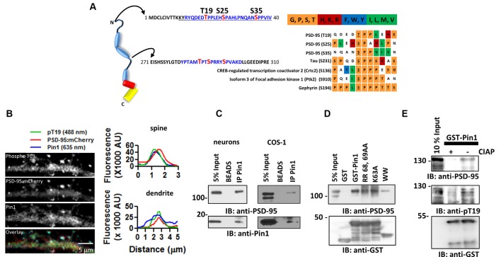
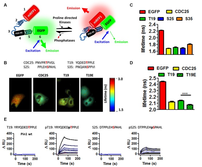
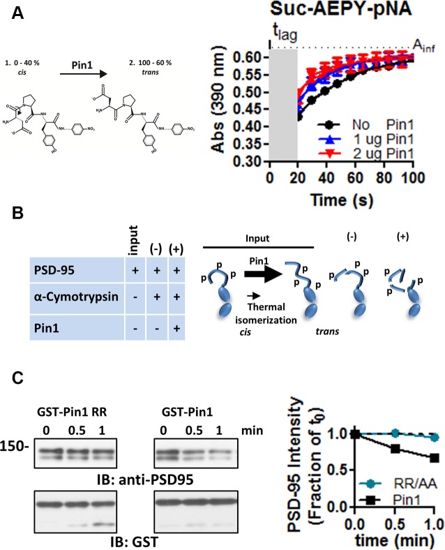
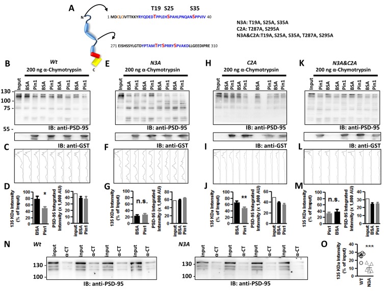
Phosphorylation-dependent peptidyl-prolyl cis-trans isomerization plays key roles in cell cycle progression, the pathogenesis of cancer, and age-related neurodegeneration. Most of our knowledge about the role of phosphorylation-dependent peptidyl-prolyl cis-trans isomerization and the enzyme catalyzing this reaction, the peptidyl-prolyl isomerase (Pin1), is largely limited to proteins not present in neurons. Only a handful of examples have shown that phosphorylation-dependent peptidyl-prolyl cis-trans isomerization, Pin1 binding, or Pin1-mediated peptidyl-prolyl cis-trans isomerization regulate proteins present at excitatory synapses. In this work, I confirm previous findings showing that Pin1 binds postsynaptic density protein-95 (PSD-95) and identify an alternative binding site in the phosphorylated N-terminus of the PSD-95. Pin1 associates via its WW domain with phosphorylated threonine (T19) and serine (S25) in the N-terminus domain of PSD-95 and this association alters the local conformation of PSD-95. Most importantly, I show that proline-directed phosphorylation of the N-terminus domain of PSD-95 alters the local conformation of this region. Therefore, proline-directed phosphorylation of the N-terminus of PSD-95, Pin1 association, and peptidyl-prolyl cis-trans isomerization may all play a role in excitatory synaptic function and synapse development.

Keywords: Pin1; cis-trans isomerization; excitatory synaptic transmission; postsynaptic density protein 95; proline-directed phosphorylation.


Figures

Figure 1
Figure 1
The peptidylprolyl cis-trans isomerase NIMA-interacting 1 (Pin1) interacts with postsynaptic density protein-95 (PSD-95) via its WW domain. (A) (Left) Schematic diagram of PSD-95 indicating putative Pin1 binding sites (shown in blue). (Right) Amino acid sequence alignment oriented around the S/T-P motifs. A chart was generated using the PRALINE algorithm. Color-code based on amino acid groups; shown above in abbreviated form. (B) Pin1 co-localizes with phosphorylated PSD-95 in vivo. Representative confocal images of hippocampal neurons transfected with PSD-95::mCherry (2nd image) and immunostained against phospho-T19 (Top) and Pin1 (3rd image) and overlay image (lower). Line scans above a dendrite and a dendritic spine (white lines) showing good co-localization on spines (top) and dendrites (bottom), right graphs. (C) PSD-95 Pin1 interaction is present in vivo. Neurons experiments performed on endogenous proteins. For COS-1 lysates were transfected with full-length PSD-95::EGFP and Pin1 and immunoprecipitated with the anti-Pin1 antibody. Complexes were subjected to Western immunoblotting with anti-PSD-95 and anti-Pin1 antibodies, n = 3. (D) Pin1 WW domain is sufficient for PSD-95 interaction. COS cells homogenate expressing PSD-95::EGFP were incubated with GST, GST-Pin1, GST-Pin RR 68, 69 AA, GST-Pin K63A or GST-Pin1 WW domain. Complexes were subjected to Western immunoblotting with anti-PSD-95 and GST antibodies, n = 4. (E) PSD-95 Pin1 interaction is regulated by phosphorylation. COS cell homogenate expressing PSD-95::EGFP were incubated with (−) or without (+) CIAP followed by incubation with GST-Pin1. Complexes were subjected to Western immunoblotting with anti-PSD-95, anti-pT19 and GST antibodies.
Figure 2
Figure 2
Pin1 WW domain binds the N-terminus domain of PSD-95. (A) EKAR constructs used to screen for interactions with phospho-sequences in PSD-95. EKAR components: 1. mRFP1, 2. Pin1-WW domain, 3. glycine linker, 4. CDC25c substrate peptide or PSD-95 phospho-peptides, 5. ERK docking domain, 6. mEGFP, red diamond represent the addition of phosphate group. (B) EGFP fluorescence lifetime images of COS cells transfected with EGFP, EKARcyto, EKARcytoT19, EKARcytoT19E. The substrate peptide sequence is shown above. (C) The WW domain binds to T19, S25, and S35. Summary plot showing the average EGFP fluorescence lifetime from fixed mounted COS cells EGFP 2.225 ± 0.078 n = 575; CDC25 1.679 ± 0.2001 n = 256; T19 1.715 ± 0.1753; S25 1.689 ± 0.196 ns n = 200; S35 1.808 ± 0.0139. Graphs show the mean ± SEM. (D) Summary plot showing the average EGFP fluorescence lifetime from live COS cell imaging showing equal affinity to CDC25 peptide EGFP 2.444 ± 0.0072 n = 136; CDC25 2.122 ± 0.12 n = 142; T19 2.143 ± 0.082; T19E 2.074 ± 0.078 ns n = 87. A Kruskal–Wallis test with Dunn’s multiple comparison test ***p < 0.0001. (E) Kinetic interaction of GST-Pin1 with phospho and non-phosphorylated peptides in the N-terminus domain of PSD-95 as visualized by SPR in a Biacore 3000 apparatus for wt GST-Pin1. The association and dissociation phases were monitored for 200 s by following the change in RU for different concentrations of GST fusion proteins in μM: 0.25, 0.125, 0.063, 0.05, 0.0313, 0.025, 0.0156, 0.01, 0.0078 for phosphorylated or non-phosphorylated peptides. The peptide sequence is shown on top.
Figure 3
Figure 3
Pin1 isomerizes full-length PSD-95. (A) A cis-trans isomerization scheme mediated by Pin1 on the succinyl-AEPY-p-nitronilide peptide. For representation, only EPY-p-nitronilide is shown. The predicted amounts of each isomer in solution (see “Materials and Methods/References” secton). The cis-trans isomerization assay to test the functionality of the purified GST-Pin1. (B) (Left) Table showing the experiment design for the α-chymotrypsin-coupled cis-trans isomerization assay. (Right) Scheme of the Pin1-mediated isomerization reaction, only N-terminus, PDZ1 and PDZ2 are shown. Input condition contains both cis and trans-N-terminus PSD-95, (−) PSD-95 product in the presence of α-chymotrypsin alone and (+) PSD-95 product in the presence of Pin1 with α-chymotrypsin. (C) Immunoblot showing the results of the in vitro α-chymotrypsin cis-trans isomerization of full-length PSD-95. Homogenates of COS cells expressing full-length PSD-95::EGFP were incubated with the isomerase dead GST-Pin RR68, 69AA or GST-Pin1 and treated with 0.1 μg of chymotrypsin. Reactions were subjected to Western immunoblotting with anti-PSD-95 and GST antibodies. (Middle) Quantification of immunoblot band intensities normalized to time 0 for the 135 kDa band (n = 1).
Figure 4
Figure 4
Phospho-T19, S25, and S35 in the N-terminus domain of PSD-95 are sites of Pin1 isomerization. (A) Schematic diagram of PSD-95 indicating putative Pin1 binding sites (shown in blue) and phosphorylated sites in red. Abbreviated mutant is shown on right including the sites mutated on each one. (B) Immunoblot showing the result of the in vitro a-chymotrypsin cis-trans isomerization of wt PSD-95, (E) the N3A, (H) the C2A, and (K) the N3A/C2A mutant. COS cells homogenates expressing wt or mutants of PSD-95::EGFP were incubated with 1 mg BSA or GST-Pin1 and subjected to Western immunoblotting with anti-PSD-95 and anti-GST antibodies. (C,F,I,L) Integrated band intensity for all the bands. Each lane corresponds to the PSD-95 immunostained column above. (D,G,J,M) Quantification of immunoblot intensities normalized to input lane value for the 135 kDa band. (D) Wt, BSA 78.78 ± 8.628% vs. Pin1 49.74 ± 5.722%; n = 9, *p < 0.05 unpaired t-test. (G) N3A, T19A, S25A, S35A PSD-95::EGFP triple mutant BSA 23.78 ± 3.246% vs. Pin1 27.44 ± 3.689%, n = 8; (J) C2A T287A, S295A PSD-95::EGFP double mutant BSA 67.38 ± 13.39% vs. Pin1 49.68 ± 10.10%, n = 8 and (M) the N3A&C2A T19A, S25A, S35, T287A, S295A PSD-95::EGFP Penta mutant BSA 34.47 ± 7.712% vs. Pin1 37.69 ± 3.426%, n = 6. *p < 0.05 and **p < 0.01 unpaired t-tests. For all (right bar graph) the difference in rates of PSD-95 degradation by Pin1 is not explained by differences in loaded protein. (N) Isomerization assay performed as before without the addition of GST-Pin1 to lysates. Cells transfected with wild type PSD-95 or the N3A. (O) Quantification of immunoblot band intensities normalized to input lane values for the 135 KDa band 25.72 ± 1.87%, n = 6 for PSD-95::EGFP and 10.99 ± 2.37%, n = 5 for PSD-95::EGFP N3A, ***p < 0.0008. Four independent pairs of reactions shown. All values are reported as mean ± SEM. n.s., not significant.

Similar articles

Cited by

References

    1. Antonelli R., De Filippo R., Middei S., Stancheva S., Pastore B., Ammassari-Teule M., et al. . (2016). Pin1 modulates the synaptic content of NMDA receptors via prolyl-isomerization of PSD-95. J. Neurosci. 36, 5437–5447. 10.1523/JNEUROSCI.3124-15.2016 - DOI - PMC - PubMed
    1. Chowdhury D., Turner M., Patriarchi T., Hergarden A. C., Anderson D., Zhang Y., et al. . (2018). Ca2+/calmodulin binding to PSD-95 mediates homeostatic synaptic scaling down. EMBO J. 37, 122–138. 10.15252/embj.201695829 - DOI - PMC - PubMed
    1. Coba M. P., Pocklington A. J., Collins M. O., Kopanitsa M. V., Uren R. T., Swamy S., et al. . (2009). Neurotransmitters drive combinatorial multistate postsynaptic density networks. Sci. Signal. 2:ra19. 10.1126/scisignal.2000102 - DOI - PMC - PubMed
    1. Colledge M., Snyder E. M., Crozier R. A., Soderling J. A., Jin Y., Langeberg L. K., et al. . (2003). Ubiquitination regulates PSD-95 degradation and AMPA receptor surface expression. Neuron 40, 595–607. 10.1016/s0896-6273(03)00687-1 - DOI - PMC - PubMed
    1. Daum S., Lücke C., Wildemann D., Schiene-Fischer C. (2007). On the benefit of bivalency in peptide ligand/pin1 interactions. J. Mol. Biol. 374, 147–161. 10.1016/j.jmb.2007.09.019 - DOI - PubMed