. 2020 Mar 18:11:218.
doi: 10.3389/fphar.2020.00218. eCollection 2020.

Hepatoprotective Effect of Cranberry Nutraceutical Extract in Non-alcoholic Fatty Liver Model in Rats: Impact on Insulin Resistance and Nrf-2 Expression

Affiliations

Hepatoprotective Effect of Cranberry Nutraceutical Extract in Non-alcoholic Fatty Liver Model in Rats: Impact on Insulin Resistance and Nrf-2 Expression

Safaa A Faheem et al. Front Pharmacol. .

Abstract

Background: Non-alcoholic fatty liver disease (NAFLD) is a pathological accumulation of triglycerides (TGs) in the hepatocyte in the absence of alcohol intake. Untreated NAFLD is expected to progress into liver fibrosis. Cranberry is rich in polyphenols with antioxidant and anti-inflammatory activities.

Hypothesis: The present study was performed to evaluate our hypothesis of the possible anti-fibrotic effect of cranberry nutraceuticals in a high fat cholesterol diet induced (HFCD)-NAFLD in rats, focusing on improving insulin sensitivity and modulating the expression of nuclear factor erythroid-2-related factor-2 (Nrf2) (a transcription factor responsible for regulating cellular redox balance).

Method: Male albino wistar rats (12 weeks) received HFCD and/or cranberry (50 and 100 mg/kg/day, three times/week) orally for 8 consecutive weeks.

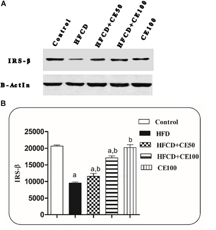
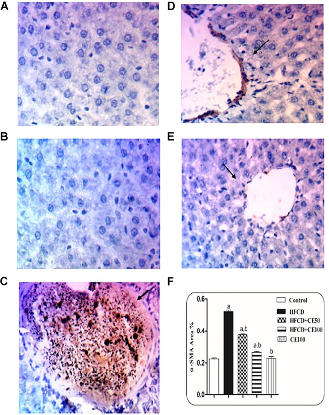
Results: In comparison to the HFCD group, cranberry treated groups (50 and 100 mg/kg) showed marked hepatoprotection, where it significantly decreased liver enzymes (alanine transaminases by 49 and 64% and aspartate transaminases by 45 and 64%; respectively), TGs, and ameliorated the histopathological alterations (such as inflammatory cells infiltration and ballooning degeneration) induced by HFCD. Cranberry also alleviated oxidative stress (malondialdehyde, glutathione, catalase and superoxide dismutase) and inflammation (tumor necrosis factor- alpha, interleukine-6 and nuclear factor kappa-b) and significantly reduced the HOMA-IR and TyG index. On the other hand, cranberry treated groups (50 and 100 mg/kg) showed a marked increase in the expression of adiponectin, by 8 and 13-fold, insulin receptor substrate-2 by 21 and 79%, and Nrf2 by 13 and 61%, respectively. Notably, cranberry significantly reduced the fibrotic markers, TGF-β and α-SMA expression and collagen deposition.

Conclusion: The present study showed that cranberry significantly attenuated NAFLD, in a dose dependent manner, which could be partially recognized by its antioxidant, anti-inflammatory activities, and its ability to improve insulin sensitivity. Notably, our study proves for the first time that the anti-fibrotic activity of cranberry is promising.

Keywords: NAFLD; Nrf-2; cranberry nutraceutical; insulin sensitivity; liver fibrosis.


Figures

FIGURE 1
FIGURE 1
UPLC-ESI-MS chromatogram of cranberry extract. (A) UPLC-ESI-MS chromatogram of cranberry extract in negative ion mode. (B) UPLC-ESI-MS chromatogram of cranberry extract in positive ion mode.
FIGURE 2
FIGURE 2
Representative photomicrographs of liver sections stained with hematoxylin and eosin (X40). (A,B) Sections taken from rat in the control and the cranberry-only groups, respectively showing normal histological structure of the central vein (CV) and surrounding hepatocyte (h). (C) Section taken from HFCD fed rats showing congestion in the portal vein (PV), fibrosis (F) with inflammatory cells infiltration (m) in the portal area associated with hyperplasia in the lining epithelium of bile duct (bd) as well as ballooning degeneration (b) and rupture (r) in the hepatocyte. (D) Section taken from HFCD rats showing lipid droplets accumulation (arrow) in the hepatocyte (X80). (E) Section taken from rats fed HFCD and co-treated with CE50 showing congestion in the portal vein (PV) and inflammatory cell infiltration (F) in the portal area as well as ballooning degeneration (b) in the hepatocyte. (F) Section taken from rats fed HFCD and co-treated with CE100 showing intact histological structure of the central vein and surrounding hepatocyte.
FIGURE 3
FIGURE 3
The effect of cranberry nutraceutical (50 and 100 mg/kg) on IRS-2 expression in HFCD-induced liver fibrosis in rats. (A) Western blot analysis of IRS-2 in rats fed HFCD and co-treated with cranberry 50 and/or cranberry 100. (B) Quantification of IRS-2 expression in rats fed HFCD and co-treated with cranberry 50 and/or cranberry 100. -Data are represented as mean ± S.E.M. (n = 5). (a) Significantly different from the control group and (b) significantly different from the HFCD groups, respectively, at p < 0.05 using ANOVA followed by Tukey–Kramer as a post hoc test.
FIGURE 4
FIGURE 4
The effect of cranberry nutraceutical (50 and 100 mg/kg) on Nrf-2 expression in HFCD-induced liver fibrosis in rats. (A) Western blot analysis of nuclear Nrf-2 in rats fed HFCD and co-treated with cranberry 50 and/or cranberry 100. (B) Quantification of the nuclear Nrf-2 expression in rats fed HFCD and co-treated with cranberry 50 and/or cranberry 100. -Data are represented as mean ± S.E.M. (n = 5). a, b: Significantly different from the control and HFCD groups, respectively, at p < 0.05 using ANOVA followed by Tukey–Kramer as a post hoc test.
FIGURE 5
FIGURE 5
The effect of cranberry nutraceutical (50 and 100 mg/kg) on TGF-β expression in HFCD-induced liver fibrosis in rats by immunohistochemical staining. (A,B) Liver sections taken from rats in the control and the cranberry-only groups showing that TGF-β expression was minimal in the hepatic tissue. (C) Sections taken from rats in the HFCD group showing extensive TGF-β expression (brown color). (D) Sections taken from rats fed HFCD and co-treated with cranberry 50 showing mild TGF-β expression. (E) Sections taken from rats fed HFCD and co-treated with cranberry 100 showing minimal TGF-β expression (brown color). All sections were magnified at (X200). (F) Quantitative image analysis expressed as percentage of area of immunopositive reaction. Data are represented as mean ± S.E.M. (n = 5). a, b: Significantly different from the control and HFCD groups, respectively, at p < 0.05 using ANOVA followed by Tukey–Kramer as a post hoc test.
FIGURE 6
FIGURE 6
The effect of cranberry nutraceutical (50 and 100 mg/kg) on α-SMA expression in HFCD-induced liver fibrosis in rats by immunohistochemical staining. (A,B) Liver sections taken from rats in the control and the cranberry-only groups showing minimal expression of α-SMA. (C) show sections taken from rats in the HFCD displaying extensive α-SMA expression (brown color). (D) Sections taken from the group fed HFCD and co-treated with cranberry 50 showing mild α-SMA expression. (E) Sections taken from rats fed HFCD and co-treated with cranberry 100 showing minimal α-SMA expression (brown color). All sections were magnified at (X200). (F) Quantitative image analysis expressed as percentage of area of immunopositive reaction. Data are represented as mean ± S.E.M. (n = 5). a, b: Significantly different from the control and HFCD groups, respectively, at p < 0.05 using ANOVA followed by Tukey–Kramer as a post hoc test.
FIGURE 7
FIGURE 7
Representative photomicrographs of liver sections stained with Masson’s trichrome. (A,B) hepatic sections taken from rat in the control and the cranberry-only groups, respectively showing normal histological structure in the portal area with minimal collagen deposition. (C) Section taken from HFCD rats showing severe congestion in the portal vein and excessive collagen fibers deposition along with pseudolobules formation in the portal area. (D) Section taken from rats fed HFCD and co-treated with cranberry 50 showing nearly an absence of pseudolobules with less collagen deposition. (E) Section taken from rats fed HFCD and co-treated with cranberry 100 showing an absence of pseudolobules and minimal collagen fibers deposition. All sections were magnified at (X40). (F) Liver hydroxyproline content in different groups, Data are represented as mean ± S.E.M. (n = 5). a, b: Significantly different from the control and HFCD groups, respectively, at p < 0.05 using ANOVA followed by Tukey–Kramer as a post hoc test.

References

    1. Ali H. A., Hussein M. A., Barakat M. A. (2015). Biochemical effects of cranberry extract in experimentally induced myocardial necrosis in rats. Benha Vet. Med. J. 28 155–162. 10.21608/bvmj.2015.32494 - DOI
    1. Angulo P. (2002). Nonalcoholic fatty liver disease. N. Engl. J. Med. 346 1221–1231. - PubMed
    1. Anhê F. F., Roy D., Pilon G., Dudonné S., Matamoros S., Varin T. V., et al. (2014). A polyphenol-rich cranberry extract protects from diet-induced obesity, insulin resistance and intestinal inflammation in association with increased Akkermansia spp. population in the gut microbiota of mice. Gut 64 872–883. 10.1136/gutjnl-2014-307142 - DOI - PubMed
    1. Anstee Q. M., Goldin R. D. (2006). Mouse models in non-alcoholic fatty liver disease and steatohepatitis research. Int. J. Exp. Pathol. 87 1–16. 10.1111/j.0959-9673.2006.00465.x - DOI - PMC - PubMed
    1. Asrih M., Jornayvaz F. R. (2014). Diets and nonalcoholic fatty liver disease: the good and the bad. Clin. Nutr. 33 186–190. 10.1016/j.clnu.2013.11.003 - DOI - PubMed

LinkOut - more resources