. 2020 Mar 17:11:327.
doi: 10.3389/fphar.2020.00327. eCollection 2020.

Functional Study of Novel Bartter's Syndrome Mutations in ClC-Kb and Rescue by the Accessory Subunit Barttin Toward Personalized Medicine

Affiliations

Functional Study of Novel Bartter's Syndrome Mutations in ClC-Kb and Rescue by the Accessory Subunit Barttin Toward Personalized Medicine

Dalila Sahbani et al. Front Pharmacol. .

Abstract

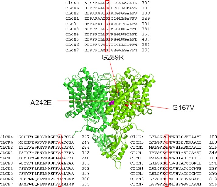
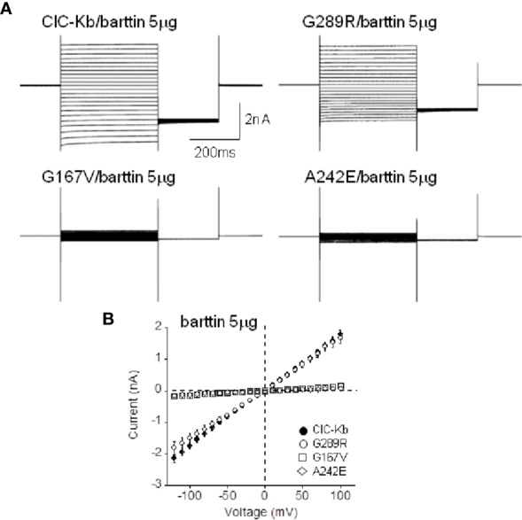
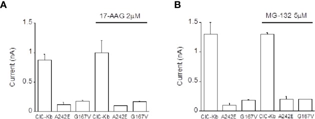
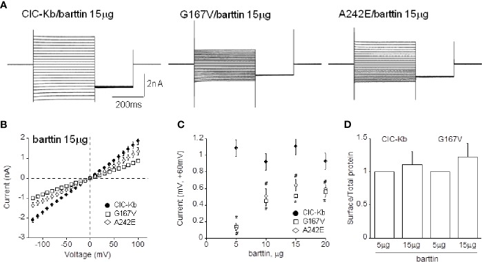
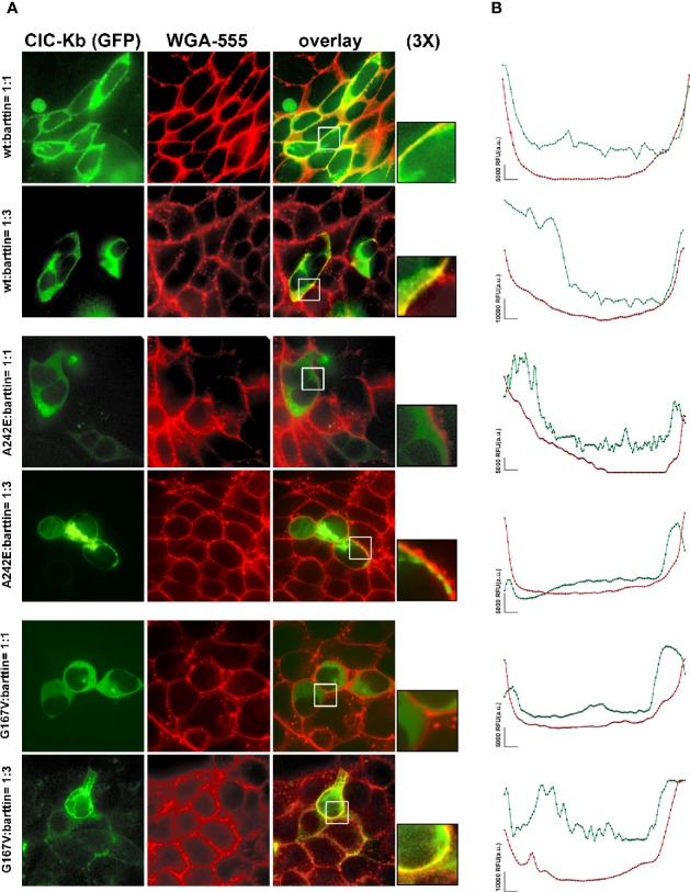
Type III and IV Bartter syndromes (BS) are rare kidney tubulopathies caused by loss-of-function mutations in the CLCNKB and BSND genes coding respectively for the ClC-Kb chloride channels and accessory subunit barttin. ClC-K channels are expressed in the Henle's loop, distal convoluted tubule, and cortical collecting ducts of the kidney and contribute to chloride absorption and urine concentration. In our Italian cohort, we identified two new mutations in CLCNKB, G167V and G289R, in children affected by BS and previously reported genetic variants, A242E, a chimeric gene and the deletion of the whole CLCNKB. All the patients had hypokalemia and metabolic alkalosis, increased serum renin and aldosterone levels and were treated with a symptomatic therapy. In order to define the molecular mechanisms responsible for BS, we co-expressed ClC-Kb wild type and channels with point mutations with barttin in HEK 293 cells and characterized chloride currents through the patch-clamp technique. In addition, we attempted to revert the functional defect caused by BS mutations through barttin overexpression. G167V and A242E channels showed a drastic current reduction compared to wild type, likely suggesting compromised expression of mutant channels at the plasma membrane. Conversely, G289R channel was similar to wild type raising the doubt that an additional mutation in another gene or other mechanisms could account for the clinical phenotype. Interestingly, increasing ClC-K/barttin ratio augmented G167V and A242E mutants' chloride current amplitudes towards wild type levels. These results confirm a genotype-phenotype correlation in BS and represent a preliminary proof of concept that molecules functioning as molecular chaperones can restore channel function in expression-defective ClC-Kb mutants.

Keywords: Bartter’s syndrome; barttin; kidney chloride channels; patch clamp; pharmacology.


Figures

Figure 1
Figure 1
Structure and alignment of ClC-Kb channel. Three dimensional representation of the ClC-Kb channel modeled upon the cryo-electron microscopy structure of the bovine ClC-K (PDB id: 5TQQ; Park et al., 2017) showing the localization of the BS mutations. The insets show the amino acids alignment of CLC proteins highlighting the position of the BS mutations.
Figure 2
Figure 2
Functional characterization of ClC-Kb mutants expressed in HEK 293 cells. (A) Representative chloride current traces of ClC-Kb wild type, G167V, G289R, and A242E channels (5 µg) co-expressed with equal amount of barttin (1:1 ratio) in HEK 293 cells. (B) IV plot showing the mean current amplitude of the indicated channels as a function of membrane potential. Data are mean ± SE; n = 11–19 cells.
Figure 3
Figure 3
Effect of Hsp90 and proteasome inhibition on ClC-Kb mutants expressed in HEK 293 cells. Bar graphs showing the effect of incubation of HEK 293 expressing ClC-Kb wild type, G167V and A242E channels with (A) 17-AAG (16 h) and with (B) MG-132 (12 h). ClC-Kb subunits were co-expressed with 5 µg of barttin. Data are mean ± SE; n = 6–9 cells.
Figure 4
Figure 4
Effect of barttin overexpression on ClC-Kb wild type, G167V and A242E current amplitude. (A) Representative chloride current traces of ClC-Kb wild type, G167V, G289R, and A242E channels (5 µg) co-expressed with 15 µg barttin (1:3 ratio) in HEK 293 cells. (B) IV plot showing the mean current amplitude as a function of membrane potential of the indicated channels co-expressed with 15 µg barttin. (C) Chloride current measured at +60 mV as a function of the amount of co-expressed barttin for ClC-Kb WT, G167V, and A242E. Data are mean ± SE; n = 6–19. p < 0.05 for G167V (*) and A242E (#) compared with the respective ClC-Kb wild type. (D) Quantification of the surface expression efficiency for ClC-Kb and G167V channels co-expressed with 5 or 15 µg of barttin calculated by dividing surface protein density to the respective total protein density. The mean surface density of ClC-Kb and G167V channels co-expressed with 15 µg of barttin was normalized to the corresponding density of the same channels co-expressed with 5 µg of barttin. Data are mean ± SE of n = 3 experiments.
Figure 5
Figure 5
Effect of barttin overexpression on ClC-Kb wild type, G167V and A242E membrane localization. (A) Immunofluorescence confocal microscopy analysis of the plasma membrane marker WGA-555 (red signal) and EGFP-ClC-Kb WT, A242E and G167V (green signal), transfected with the same amount of barttin (5 µg: 5 µg, 1:1 ratio, upper panels) or overexpressing barttin (5 µg: 15 µg, 1:3 ratio, lower panels) in HEK 293 cells grown on coverslips. The overlay column reports the co-localization of the two fluorescence signals. Areas indicated by the white boxes are magnified three times. (B) Representative quantitative analysis of EGFP-ClC-Kb WT, A242E and G167V (green signal), and WGA (red signal) fluorescence distribution between plasma membrane and cytosol along a fixed yellow line. Peaks of ClC-Kb overlapped with peaks of WGA in cells transfected with either ClC-Kb WT or with both A242E and G167V mutants co-transfected with an excess of barttin. Similar results were obtained when the analysis was repeated in 10 transfected cells in four different fields using MetaMorph as imaging software. The vertical bar indicates 5,000 RFU (a.u.). WGA, Wheat Germ Agglutinin.

Similar articles

Cited by

References

    1. Alhammadi A. H., Khalifa M., Alnaimi L. (2014). An infant with poor weight gain and hypochloremic metabolic alkalosis: a case report. Int. J. Gen. Med. 7, 389–391. 10.2147/IJGM.S66550 - DOI - PMC - PubMed
    1. Andrini O., Keck M., L’Hoste S., Briones R., Mansour-Hendili L., Grand T., et al. (2014). CLCNKB mutations causing mild Bartter syndrome profoundly alter the pH and Ca2+ dependence of ClC-Kb channels. Pflugers Arch. 466 (9), 1713–1723. 10.1007/s00424-013-1401-2 - DOI - PubMed
    1. Andrini O., Keck M., Briones R., Lourdel S., Vargas-Poussou R., Teulon J. (2015). ClC-K chloride channels: emerging pathophysiology of Bartters yndrome type3. Am. J. Physiol. Renal Physiol. 308, F1324–F1334. 10.1152/ajprenal.00004.2015 - DOI - PubMed
    1. Barrallo-Gimeno A., Gradogna A., Zanardi I., Pusch M., Estévez R. (2015). Regulatory/Auxiliary subunits of CLC chloride channel/transport proteins. J. Physiol. 593 (18), 4111–4127. 10.1113/JP270057 - DOI - PMC - PubMed
    1. Bettinelli A., Borsa N., Bellantuono R., Syrèn M. L., Calabrese R., Edefonti A., et al. (2007). Patients with biallelic mutations in the chloride channel gene CLCNKB: long-term management and outcome. Am. J. Kidney Dis. 49 (1), 91–98. 10.1053/j.ajkd.2006.10.001 - DOI - PubMed