. 2020 Mar 17:11:189.
doi: 10.3389/fimmu.2020.00189. eCollection 2020.

Chronic Intra-Uterine Ureaplasma parvum Infection Induces Injury of the Enteric Nervous System in Ovine Fetuses

Affiliations

Chronic Intra-Uterine Ureaplasma parvum Infection Induces Injury of the Enteric Nervous System in Ovine Fetuses

Cathelijne Heymans et al. Front Immunol. .

Erratum in

Abstract

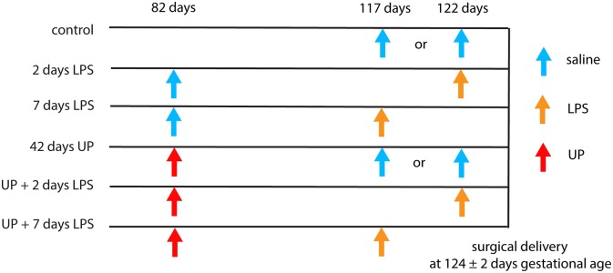
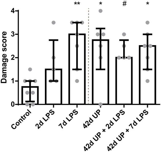
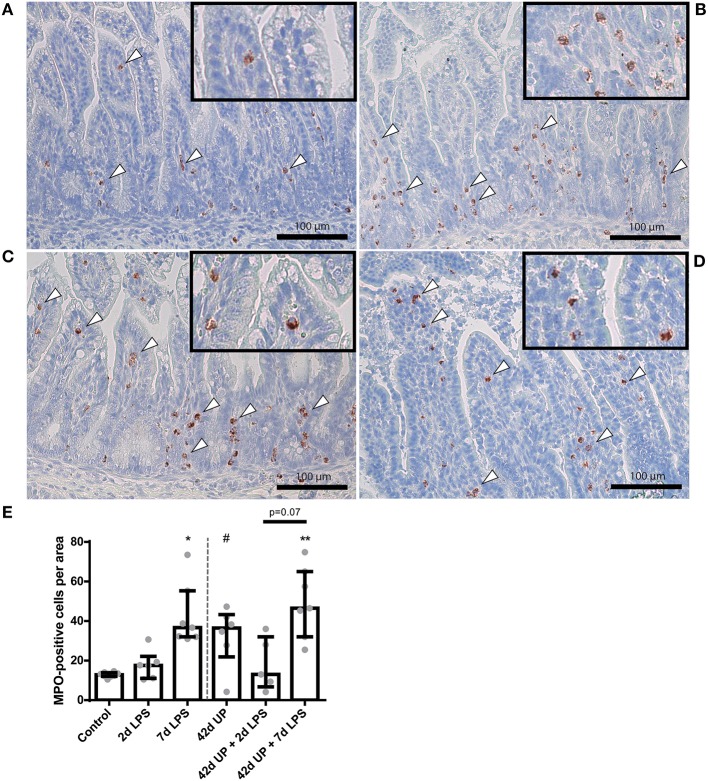
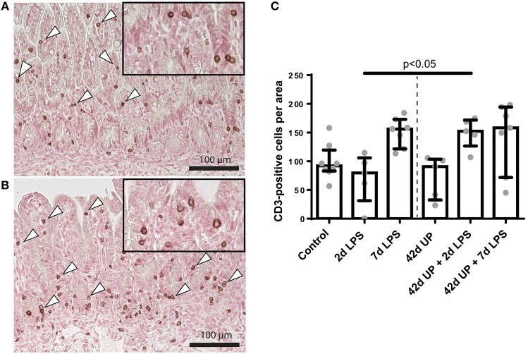
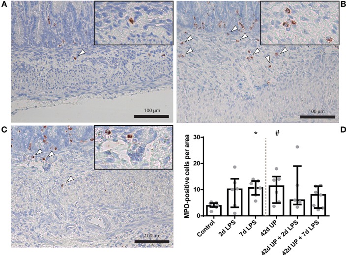
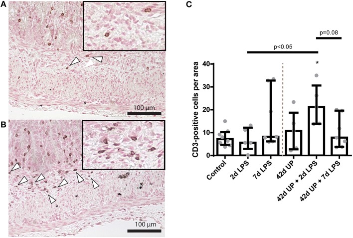
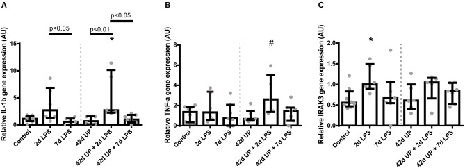
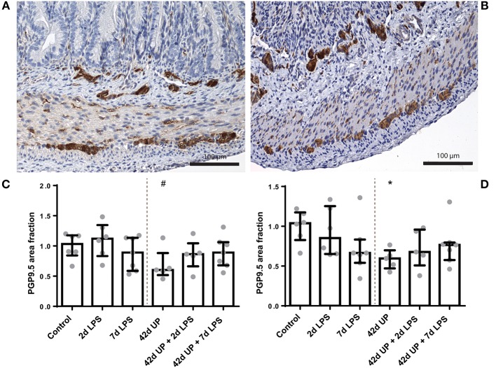
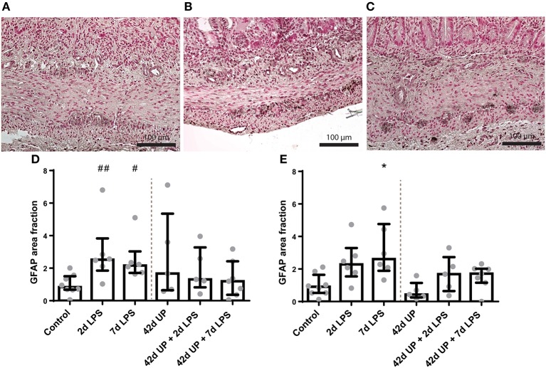
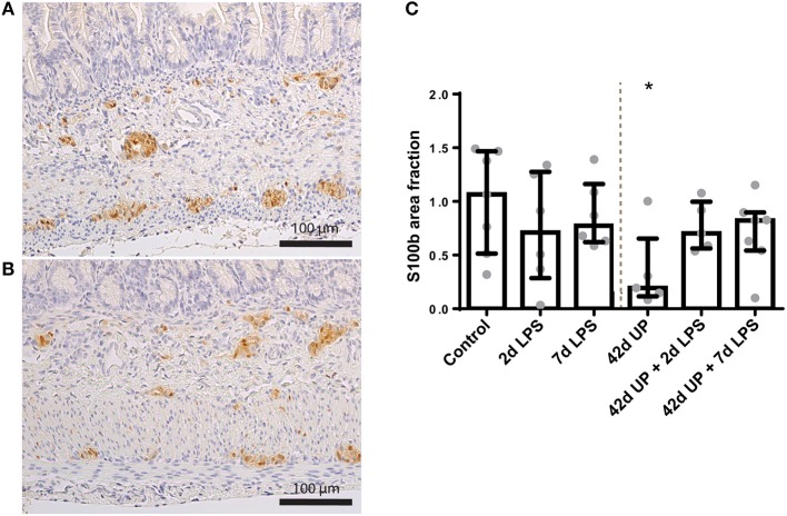
Background: Chorioamnionitis, inflammation of the fetal membranes during pregnancy, is often caused by intra-amniotic (IA) infection with single or multiple microbes. Chorioamnionitis can be either acute or chronic and is associated with adverse postnatal outcomes of the intestine, including necrotizing enterocolitis (NEC). Neonates with NEC have structural and functional damage to the intestinal mucosa and the enteric nervous system (ENS), with loss of enteric neurons and glial cells. Yet, the impact of acute, chronic, or repetitive antenatal inflammatory stimuli on the development of the intestinal mucosa and ENS has not been studied. The aim of this study was therefore to investigate the effect of acute, chronic, and repetitive microbial exposure on the intestinal mucosa, submucosa and ENS in premature lambs. Materials and Methods: A sheep model of pregnancy was used in which the ileal mucosa, submucosa, and ENS were assessed following IA exposure to lipopolysaccharide (LPS) for 2 or 7 days (acute), Ureaplasma parvum (UP) for 42 days (chronic), or repetitive microbial exposure (42 days UP with 2 or 7 days LPS). Results: IA LPS exposure for 7 days or IA UP exposure for 42 days caused intestinal injury and inflammation in the mucosal and submucosal layers of the gut. Repetitive microbial exposure did not further aggravate injury of the terminal ileum. Chronic IA UP exposure caused significant structural ENS alterations characterized by loss of PGP9.5 and S100β immunoreactivity, whereas these changes were not found after re-exposure of chronic UP-exposed fetuses to LPS for 2 or 7 days. Conclusion: The in utero loss of PGP9.5 and S100β immunoreactivity following chronic UP exposure corresponds with intestinal changes in neonates with NEC and may therefore form a novel mechanistic explanation for the association of chorioamnionitis and NEC.

Keywords: Ureaplasma parvum; chorioamnionitis; enteric nervous system; intra-amniotic infection; necrotizing enterocolitis; preterm birth; sheep.


Figures

Figure 1
Figure 1
Different intervention study groups. All injections were delivered by ultrasound-guided amniocentesis. Timing is shown in gestational days.
Figure 2
Figure 2
Increased mucosal injury in all groups, except for the animals exposed to 2 days LPS. #p = 0.06, *p < 0.05, **p < 0.005 compared to the control.
Figure 3
Figure 3
Representative images of mucosal neutrophil influx reflected by myeloperoxidase (MPO)-positive cell (indicated by white triangles) counts of the control (A), 7 days lipopolysaccharide (LPS) (B), Ureaplasma parvum (UP) (C), and UP prior to 7 days LPS groups (D). (E) Increased MPO count in animals exposed to 7 days LPS, UP, and UP prior to 7 days LPS. #p = 0.08, *p < 0.01, **p < 0.005 compared to the control.
Figure 4
Figure 4
Representative images of mucosal T cell influx reflected by CD3-positive cell (indicated by white triangles) counts of the control (A) and Ureaplasma parvum (UP) prior to 2 days lipopolysaccharide (LPS) (B). (C) Increased CD3 count in animals exposed to UP prior to 2 days LPS compared to the 2 days LPS group.
Figure 5
Figure 5
Representative images of submucosal neutrophil influx reflected by myeloperoxidase (MPO)-positive cell (indicated by white triangles) counts of the control (A), 7 days lipopolysaccharide (LPS) (B) and Ureaplasma parvum (UP) (C). (D) Increased MPO count in animals exposed to 7 days LPS and UP. #p = 0.06, *p < 0.05 compared to the control.
Figure 6
Figure 6
Representative images of submucosal T cell influx reflected by CD3-positive cell (indicated by white triangles) counts of the control (A) and Ureaplasma parvum (UP) prior to 2 days lipopolysaccharide (LPS) groups (B). (C) Increased CD3 count in animals exposed to UP prior to 2 days LPS. *p < 0.05 compared to the control.
Figure 7
Figure 7
Relative mRNA levels of IL-1β (A), TNF-α (B), and IRAK3 (C) in arbitrary unit (AU). (A) Increased IL-1β mRNA level in animals exposed to 2 days lipopolysaccharide (LPS) and Ureaplasma parvum (UP) prior to 2 days LPS. *p < 0.05 compared to the control. (B) Increased TNF-α mRNA level in animals exposed to UP prior to 2 days LPS. #p = 0.07 compared to the control. (C) Increased IRAK3 mRNA level in animals exposed to 2 days LPS. *p < 0.05 compared to the control.
Figure 8
Figure 8
Representative images of PGP9.5 immunoreactivity in the submucosal and myenteric plexus of the control (A) and Ureaplasma parvum (UP) (B) groups. Area fraction of PGP9.5 in the submucosal plexus (C) and myenteric plexus (D) as fold increase over the control value. (C) PGP9.5-positive surface area tended to be decreased in the submucosal plexus of animals exposed to UP. #p = 0.08 compared to the control. (D) Decreased PGP9.5-positive surface area in the myenteric plexus of animals exposed to UP. *p < 0.05 compared to the control.
Figure 9
Figure 9
Representative images of glial fibrillary acidic protein (GFAP) immunoreactivity in the submucosal and myenteric plexus of the control (A), 2 days lipopolysaccharide (LPS) (B), and 7 days LPS (C) group. Area fraction of GFAP in the submucosal plexus (D) and myenteric plexus (E) as fold increase over the control value. (D) The GFAP-positive surface area tended to be increased in the submucosal plexus of animals exposed to 2- and 7 days LPS. ##p = 0.07, #p = 0.09 compared to the control. (E) Increased GFAP-positive surface area in the myenteric plexus of animals exposed to 7 days LPS. *p < 0.05 compared to the control.
Figure 10
Figure 10
Representative images of S100β immunoreactivity in the submucosal and myenteric plexus of the control (A) and Ureaplasma parvum (UP) group (B). Area fraction of S100β in the myenteric plexus (C) as fold increase over the control value. (C) The S100β-positive surface area was decreased in the myenteric plexus of animals exposed to UP. *p < 0.05 compared to the control.

Similar articles

Cited by

References

    1. Murphy SL, Mathews TJ, Martin JA, Minkovitz CS, Strobino DM. Annual Summary of Vital Statistics: 2013–2014. Pediatrics. (2017) 139:e20163239. 10.1542/peds.2016-3239 - DOI - PubMed
    1. Howson CP, Kinney MV, McDougall L, Lawn JE, Born Too Soon Preterm Birth Action Group . Born too soon: preterm birth matters. Reprod Health. (2013) 10(Suppl. 1):S1. 10.1186/1742-4755-10-S1-S1 - DOI - PMC - PubMed
    1. Blencowe H, Cousens S. Addressing the challenge of neonatal mortality. Trop Med Int Health. (2013) 18:303–12. 10.1111/tmi.12048 - DOI - PubMed
    1. Blencowe H, Cousens S, Oestergaard MZ, Chou D, Moller AB, Narwal R, et al. . National, regional, and worldwide estimates of preterm birth rates in the year 2010 with time trends since 1990 for selected countries: a systematic analysis and implications. Lancet. (2012) 379:2162–72. 10.1016/S0140-6736(12)60820-4 - DOI - PubMed
    1. Galinsky R, Polglase GR, Hooper SB, Black MJ, Moss TJ. The consequences of chorioamnionitis: preterm birth and effects on development. J Pregnancy. (2013) 2013:412831. 10.1155/2013/412831 - DOI - PMC - PubMed

Publication types

MeSH terms

Substances

LinkOut - more resources