. 2020 Mar 11:2020:8681361.
doi: 10.1155/2020/8681361. eCollection 2020.

Long Noncoding RNA Lnc-TLN2-4:1 Suppresses Gastric Cancer Metastasis and Is Associated with Patient Survival

Affiliations

Long Noncoding RNA Lnc-TLN2-4:1 Suppresses Gastric Cancer Metastasis and Is Associated with Patient Survival

Yuyun Wu et al. J Oncol. .

Abstract

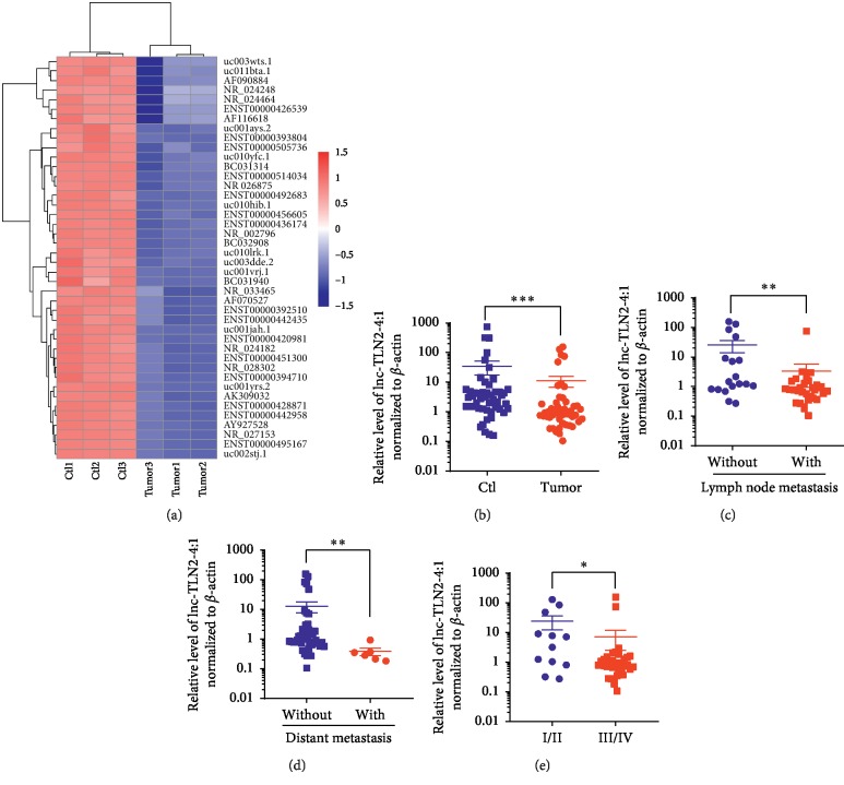
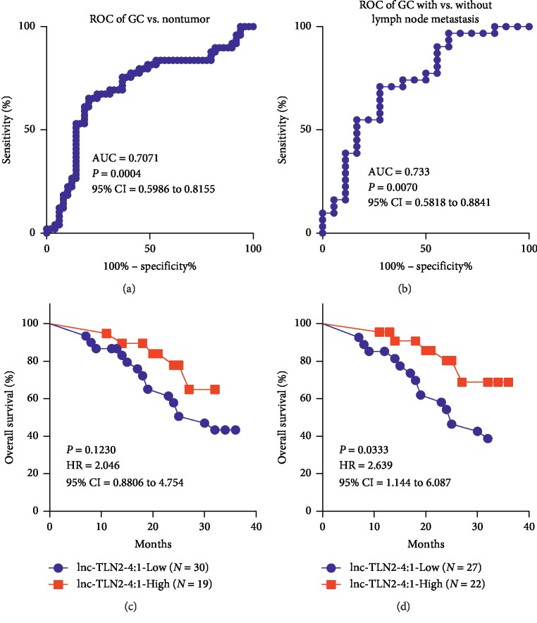
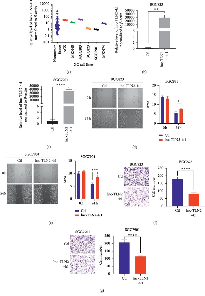
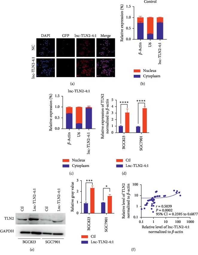
Gastric cancer (GC) is one of the most common malignancies worldwide, and the tumor metastasis leads to poor outcomes of GC patients. Long noncoding RNAs (lncRNAs) have emerged as new regulatory molecules that play a crucial role in tumor metastasis. However, the biological function and underlying mechanism of numerous lncRNAs in GC metastasis remain largely unclear. Here, we report a novel lncRNA, lnc-TLN2-4:1, whose expression is decreased in GC tissue versus matched normal tissue, and its low expression is involved in the lymph node and distant metastases of GC, as well as poor overall survival rates of GC patients. We further found that lnc-TLN2-4:1 inhibits the ability of GC cells to migrate and invade but does not influence GC cell proliferation and confirmed that lnc-TLN2-4:1 is mainly located in the cytoplasm of GC cells. We then found that lnc-TLN2-4:1 increases the mRNA and protein expression of TLN2 in GC cells and there is a positive correlation between the expression of lnc-TLN2-4:1 and TLN2 mRNA in GC tissue. Collectively, we identified a novel lncRNA, lnc-TLN2-4:1, in GC, where lnc-TLN2-4:1 represses cell migration and invasion. The low expression of lnc-TLN2-4:1 is associated with poor overall survival rates of GC patients. These suggest that lnc-TLN2-4:1 may be a tumor suppressor during GC metastasis.


Conflict of interest statement

The authors declare that they have no conflicts of interest.

Figures

Figure 1
Figure 1
Lnc-TLN2-4:1 expression is frequently decreased in GC tissues compared with matched normal tissues and is associated with GC metastasis. (a) A heatmap shows the aberrant expression of lncRNAs in three pairs of GC and matched normal tissues detected by a Human LncRNA Microarray. (b) Scatter plots show the expression of lnc-TLN2-4:1 in 49 pairs of GC and matched normal tissues, detected by qRT-PCR, and β-actin serves as the internal control. (c) Scatter plots show the expression of lnc-TLN2-4:1 in 49 GC tissues, 31 of which are of lymph node metastasis and 18 of which are not. (d) Scatter plots show the expression of lnc-TLN2-4:1 in 49 GC tissues, 6 of which are of distant metastasis and 43 of which are not. (e) Scatter plots show the expression of lnc-TLN2-4:1 in 49 GC tissues, 12 of which are the sum of TNM stage I and II, and 37 of which are the sum of TNM stage III and IV. P < 0.05, ∗∗P < 0.01, and ∗∗∗P < 0.0001.
Figure 2
Figure 2
Lnc-TLN2-4:1 expression is associated with overall survival rates of GC patients. (a) ROC curve shows that the expression of lnc-TLN2 has an AUC of 0.7071 in distinguishing GC tissue from nontumor tissue, with the sensitivity of 65.31% and the specificity of 81.25% and the cutoff value of 1.246. (b) ROC curve shows that the expression of lnc-TLN2 has an AUC of 0.733 in distinguishing GC tissue from nontumor tissue, with the sensitivity of 70.97% and the specificity of 72.22% and the cutoff value of 1.016. (c) Overall survival analysis shows the survival rates of GC patients with low or high expression of lnc-TLN2-4:1, which is defined by the cutoff value of 1.246. (d) Overall survival analysis shows the survival rates of GC patients with low or high expression of lnc-TLN2-4:1, which is defined by the cutoff value of 1.016.
Figure 3
Figure 3
Lnc-TLN2-4:1 represses GC cell migration and invasion in vitro. (a) Scatter plots show the expression of lnc-TN2-4:1 in 49 nontumor tissues and GC cell lines, detected by qRT-PCR, and β-actin serves as the internal control. (b and c) Bars show the expression of lnc-TLN2-4:1 in BGC823 and SGC7901 cells which were transfected with lnc-TLN2-4:1-overexpressing vectors, detected by qRT-PCR, and β-actin serves as the internal control. (d and e) Wound healing experiments show the abilities of BGC823 and SGC7901 cells which were transfected with lnc-TLN2-4:1-overexpressing vectors to migrate. Bars show the statistics based on three independent experiments. Area indicates the area without cells in the images, calculated by Image J. (f and g) Transwell experiments show the ability of BGC823 and SGC7901 cells which were transfected with lnc-TLN2-4:1-overexpressing vectors to invasive. Bars show the statistics based on three independent experiments. P < 0.05, ∗∗P < 0.01, ∗∗∗P < 0.001, and ∗∗∗∗P < 0.0001.
Figure 4
Figure 4
Lnc-TLN2-4:1 is located in the GC cell cytoplasm, and its expression is positively correlated with TLN2 expression in GC tissues. (a) Fluorescence in situ hybridization (FISH) shows the expression and location of lnc-TLN2-4:1 in BGC823 cells. DAPI indicates cell nucleus. GFP indicates the expression status of the vectors (negative control and lnc-TLN2-4:1-overexpressing vectors). Red fluorescence indicates the expression of lnc-TLN2-4:1. (b and c) Bars show the relative expression % of lnc-TLN2-4:1 in the cytoplasm and nucleus of BGC823 cells which were transfected with the control or lnc-TLN2-4:1-overexpressing vectors, detected by qRT-PCR, and β-actin serves as the internal control in the cytoplasm and U6 serves as the internal control in the nucleus. (d) Bars show the expression of TLN2 in BGC823 and SGC7901 cells which were transfected with control or lnc-TLN2-4:1-overexpressing vectors, detected by qRT-PCR, and β-actin serves as the internal control. (e) Western blotting shows the protein expression of TLN2 in BGC823 and SGC7901 cells which were transfected with the control or lnc-TLN2-4:1-overexpressing vectors, and GAPDH serves as the internal control. Bars show the statistics based three independent experiments. (f) The correlation between the expression of TLN2 mRNA and lnc-TLN2-4:1 in 49 pairs of GC tissues, detected by qRT-PCR, and β-actin serves as the internal control. P < 0.05, ∗∗∗P < 0.001, and ∗∗∗∗P < 0.0001.

Similar articles

Cited by

References

    1. Bray F., Ferlay J., Soerjomataram I., Siegel R. L., Torre L. A., Jemal A. Global cancer statistics 2018: GLOBOCAN estimates of incidence and mortality worldwide for 36 cancers in 185 countries. CA: A Cancer Journal for Clinicians. 2018;68(6):394–424. doi: 10.3322/caac.21492. - DOI - PubMed
    1. Salati M., Orsi G., Smyth E., et al. Gastric cancer: translating novels concepts into clinical practice. Cancer Treatment Reviews. 2019;79 doi: 10.1016/j.ctrv.2019.101889.101889 - DOI - PubMed
    1. Alyami M., Hübner M., Grass F., et al. Pressurised intraperitoneal aerosol chemotherapy: rationale, evidence, and potential indications. The Lancet Oncology. 2019;20(7):e368–e377. doi: 10.1016/s1470-2045(19)30318-3. - DOI - PubMed
    1. Zeng S., Xiao Y. F., Tang B., et al. Long noncoding RNA in digestive tract cancers: function, mechanism, and potential biomarker. The Oncologist. 2015;20(8):898–906. doi: 10.1634/theoncologist.2014-0475. - DOI - PMC - PubMed
    1. Kopp F., Mendell J. T. Functional classification and experimental dissection of long noncoding RNAs. Cell. 2018;172(3):393–407. doi: 10.1016/j.cell.2018.01.011. - DOI - PMC - PubMed

LinkOut - more resources