. 2020 Mar 16:2020:3495836.
doi: 10.1155/2020/3495836. eCollection 2020.

Electroacupuncture Ameliorates Cerebral I/R-Induced Inflammation through DOR-BDNF/TrkB Pathway

Affiliations

Electroacupuncture Ameliorates Cerebral I/R-Induced Inflammation through DOR-BDNF/TrkB Pathway

Yue Geng et al. Evid Based Complement Alternat Med. .

Abstract

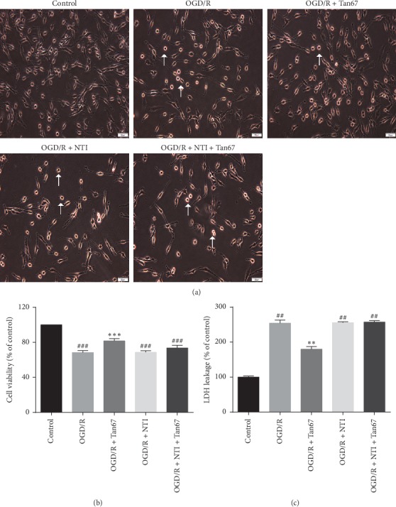
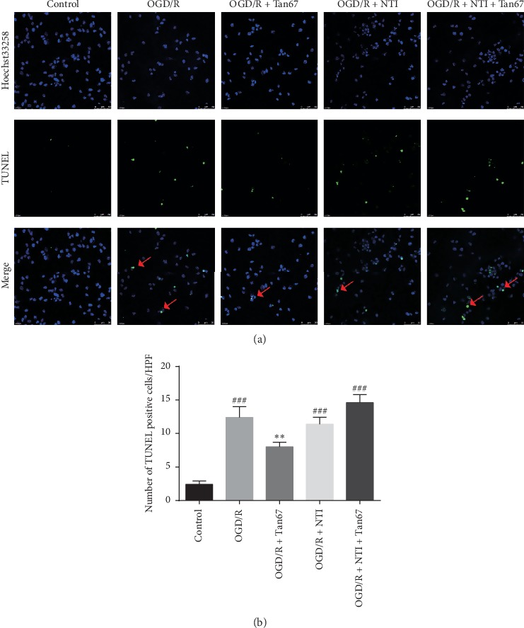
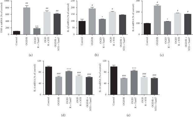
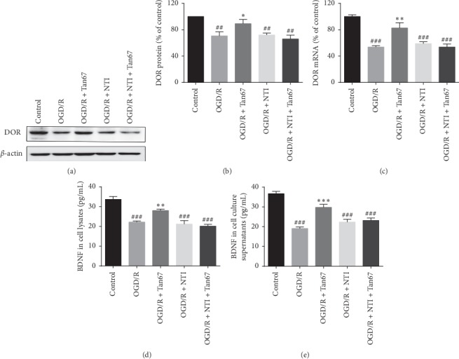
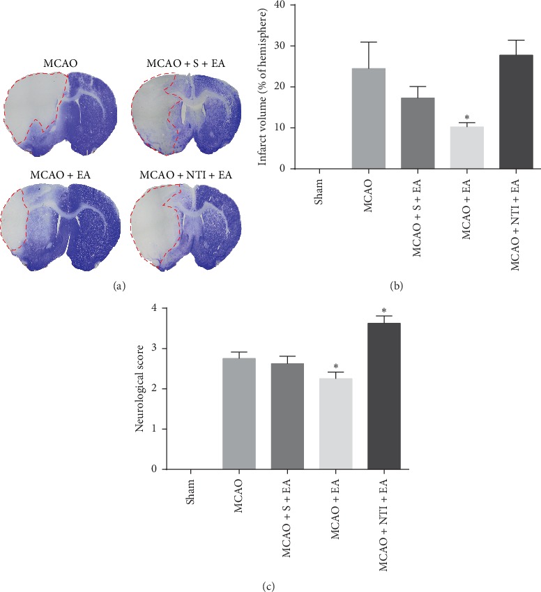
The beneficial effects of electroacupuncture (EA) at Shuigou (GV26) and Neiguan (PC6) on poststroke rehabilitation are critically related to the activation of the delta-opioid receptor (DOR). The underlying anti-inflammatory mechanisms in DOR activation and EA-mediated neuroprotection in cerebral ischemia/reperfusion (I/R) injury were investigated in the current study. Cell proliferation and apoptosis were detected by morphological changes, cell counting kit-8 (CCK-8) assay, lactate dehydrogenase (LDH) release, and TUNEL staining. The mRNA levels were evaluated by using real-time quantitative polymerase chain reaction (RT-qPCR), and the protein expression was measured by western blot or enzyme-linked immunosorbent assay (ELISA) in vitro. Infarct volume was examined by cresyl violet (CV) staining, neurologic recovery was assessed by neurological deficit scores, and pro- and anti-inflammatory cytokines were determined by immunofluorescence in vivo. DOR activation greatly ameliorated morphological injury, reduced LDH leakage and apoptosis, and increased cell viability. It reversed the oxygen-glucose deprivation/reoxygenation- (OGD/R-) induced downregulation of DOR mRNA and protein, as well as BDNF protein. DOR activation also reduced proinflammatory cytokine gene expression, including TNF-α, IL-1β, and IL-6, and at the same time, increased anti-inflammatory cytokines IL-4 and IL-10 in OGD/R challenged PC12 cells. EA significantly reduced middle cerebral artery occlusion/reperfusion- (MCAO/R-) induced infarct volume and attenuated neurologic deficit scores. It markedly increased the expression of IL-10 and decreased IL-1β, while sham EA did not have any protective effect in MCAO/R-injured rats. DOR activation plays an important role in neuroprotection against OGD/R injury by inhibiting inflammation via the brain-derived neurotrophic factor/tropomyosin-related kinase B (BDNF/TrkB) pathway. The neuroprotective efficacy of EA at Shuigou (GV26) and Neiguan (PC6) on cerebral I/R injury may be also related to the inhibition of inflammatory response through the DOR-BDNF/TrkB pathway.


Conflict of interest statement

The authors declare that they have no conflicts of interest.

Figures

Figure 1
Figure 1
DOR activation increases cell viability in OGD/R-exposed PC12 cells. The cellular morphological changes, cell viability, and LDH level were evaluated after exposure to 6 h OGD followed by 24 h reperfusion (OGD/R). The DOR agonist or antagonist treatment is 30 min before OGD. In the coadministration group, antagonist NTI was added to the cell culture medium 30 min before the agonist Tan67 incubation. (a) Representative images of PC12 cells in the sham group, OGD/R group, agonist group, antagonist group, and coadministration group. Cell viability was measured by (b) CCK-8 assay and (c) LDH release. Activation of DOR attenuated the PC12 cells injury, whereas it was totally abolished by preincubation of DOR antagonist. Scale bar = 50 μm. N = 6 in each group. ###P < 0.001 and ##P < 0.01 vs. control group. ∗∗∗P < 0.001 and ∗∗P < 0.01 vs. OGD/R group.
Figure 2
Figure 2
DOR activation protects against OGD/R-induced cell apoptosis. The apoptotic cells were evaluated after exposure to 6 h OGD followed by 24 h reperfusion (OGD/R). (a) The top panels are representative images of TUNEL staining (400x) in indicated groups. Red arrows indicate colocalization (yellow) between TUNEL-positive apoptotic cells (green) and cell nuclei (blue) in PC12 cells. Cell nuclei were visualized by Hoechst 33258 staining (blue). (b) The bottom panel is the statistical analysis of the number of apoptotic cells. DOR activation reduced cell apoptosis, and NTI blocked the effect. Scale bar = 50 μm. N = 3 in each group. ###P < 0.001 vs. control group and ∗∗P < 0.01 vs. OGD/R group.
Figure 3
Figure 3
DOR activation reduces inflammatory response. The expression of proinflammatory cytokines (e.g., TNF-α, IL-1β, and IL-6) and anti-inflammatory cytokines (e.g., IL-4 and IL-10) was evaluated by RT-qPCR at 24 h of reperfusion following 6 h OGD. Statistical analysis of (a) TNF-α, (b) IL-1β, (c) IL-6, (d) IL-4, and (e) IL-10 mRNA expression is shown. DOR activation prevented the OGD/R-induced upregulation of proinflammatory factors (TNF-α, IL-1β, and IL-6) and downregulation of the anti-inflammatory factors (IL-4 and IL-10), and the effects were blocked by NTI treatment. N = 3 in each group. #P < 0.05 and ###P < 0.001 vs. control group. P < 0.05, ∗∗P < 0.01, and ∗∗∗P < 0.001 vs. OGD/R group.
Figure 4
Figure 4
DOR activation elevates DOR and BDNF expression. The mRNA level of DOR was determined by RT-qPCR, and protein expression of DOR and BDNF was measured by western blot or ELISA at 24 h of reperfusion following 6 h OGD. (a) Representative western blot images of DOR expression. (b) Statistical analysis of DOR protein expression. (c) Statistical analysis of DOR mRNA expression. (d) Statistical analysis of BDNF protein expression in cell lysates. (e) Statistical analysis of BDNF protein expression in cell culture supernatants. DOR activation inhibited the OGD-induced decline of DOR mRNA, DOR protein, and BDNF protein expression in PC12 cells, while NTI blocked the effect. N = 3 in each group. #P < 0.05 and ###P < 0.001 vs. control group. P < 0.05, ∗∗P < 0.01, and ∗∗∗P < 0.001 vs. OGD/R group.
Figure 5
Figure 5
EA-mediated neuroprotective effects in infarct volume and neurologic deficit scores are reversed by DOR antagonist. Infarct volume and neurologic deficit scores were evaluated at 24 h of reperfusion following 90 min MCAO. (a) Representative images of brain infarct volume presented typically in the striatum and cortex indicated by CV staining. The area of pallor, which is inside the red circle, delineates the ischemic core. (b) Statistical analysis of the infarct volume in each group. (c) Statistical analysis of the neurological deficits scores in each group. DOR antagonist NTI abolished the reduction on infarct volume and neurologic deficit scores by EA stimulation at Shuigou (GV26) and Neiguan (PC6). N = 8 in each group. P < 0.05 vs. MCAO group.
Figure 6
Figure 6
DOR antagonist reversed EA-induced neuroprotective effects in inflammatory responses. Proinflammatory cytokines (IL-1β) and anti-inflammatory cytokines (IL-10) were evaluated at 24 h of reperfusion following 90 min MCAO. (a) Representative immunofluorescent staining images of IL-1β positive cells in the cortex. (b) Representative images of IL-10 positive immunofluorescent staining in the cortex. (c) Statistical analysis of the percentage of IL-1β positive cells. (d) Statistical analysis of the mean density of IL-10 positive staining. Green arrows indicate colocalization (pink) between IL-1β positive cells (red) and cell nuclei (blue). Red arrows indicate that IL-10 positive staining (green) is found diffusely distributed across the cytosol and located around the nuclei (blue). Cell nuclei were visualized by Hoechst 33258 staining (blue). EA at Shuigou (GV26) and Neiguan (PC6) reduces the inflammatory factor IL-1β and increases the anti-inflammatory factor IL-10, and NTI reversed the EA effect. Scale bar = 50 μm. N = 4 in each group. ###P < 0.001 vs. sham group. ∗∗P < 0.01 vs. MCAO group.

References

    1. Wang W., Jiang B., Sun H., et al. Prevalence, incidence, and mortality of stroke in China: results from a nationwide population-based survey of 480 687 adults. Circulation. 2017;135(8):759–771. doi: 10.1161/circulationaha.116.025250. - DOI - PubMed
    1. Koh S.-H., Park H.-H. Neurogenesis in stroke recovery. Translational Stroke Research. 2017;8(1):3–13. doi: 10.1007/s12975-016-0460-z. - DOI - PubMed
    1. Wang P., He Y., Li D., et al. Class I PI3K inhibitor ZSTK474 mediates a shift in microglial/macrophage phenotype and inhibits inflammatory response in mice with cerebral ischemia/reperfusion injury. Journal of Neuroinflammation. 2016;13(1):p. 192. doi: 10.1186/s12974-016-0660-1. - DOI - PMC - PubMed
    1. Shen M.-H., Zhang C.-B., Zhang J.-H., Li P.-F. Electroacupuncture attenuates cerebral ischemia and reperfusion injury in middle cerebral artery occlusion of rat via modulation of apoptosis, inflammation, oxidative stress, and excitotoxicity. Evidence-Based Complementary and Alternative Medicine. 2016;2016:15. doi: 10.1155/2016/9438650.9438650 - DOI - PMC - PubMed
    1. Liu A.-J., Li J.-H., Li H.-Q., et al. Electroacupuncture for acute ischemic stroke: a meta-analysis of randomized controlled trials. The American Journal of Chinese Medicine. 2015;43(8):1541–1566. doi: 10.1142/s0192415x15500883. - DOI - PubMed

LinkOut - more resources