

Predictive value of p16INK4a, Ki-67 and ProExC immuno-qualitative features in LSIL progression into HSIL
- PMID: 32256722
- PMCID: PMC7086290
- DOI: 10.3892/etm.2020.8496
Predictive value of p16INK4a, Ki-67 and ProExC immuno-qualitative features in LSIL progression into HSIL
Abstract
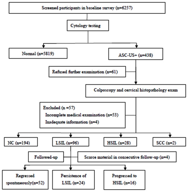
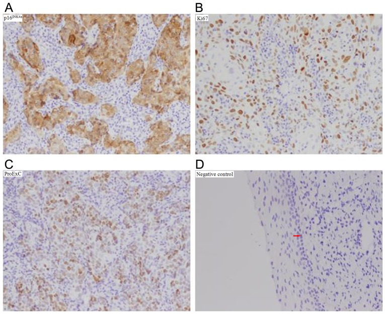
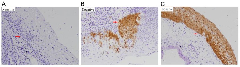
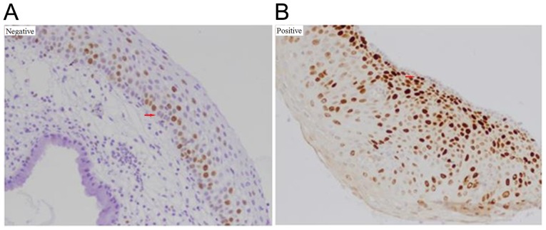
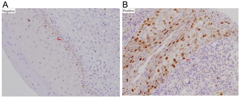
The current nested case-control study was conducted to explore the prognostic value of cyclin-dependent kinase inhibitor 2A (p16INK4a), marker of proliferation Ki-67 (Ki-67) and immunohistochemical cocktail containing antibodies directed against topoisomerase IIα (TOP2A) and minichromosome maintenance 2 (MCM2) proteins (ProExC) immuno-qualitative features to predict low-grade squamous intraepithelial lesion (LSIL) progression. A total of 92 LSIL patients were followed-up for 2 years, where those with high-grade squamous intraepithelial lesion (HSIL) or persistent LSIL were designated as the case group and those who spontaneously regressed were designated as the control group. The infection status of human papillomavirus (HPV) was evaluated using flow-through hybridization and gene chip, whilst the expression of p16INK4a, Ki-67 and ProExC were tested in LSIL patient biopsies by immunohistochemistry. All data were collected at the beginning of the follow-up and patient outcomes were diagnosed by histopathological examination. To analyze the risk factors for LSIL progression, sensitivity, specificity, positive-negative predictive value (PPV-NPV), positive-negative likelihood ratio (PLR-NLR), Youden's index (YI) and multinomial logistic regression analysis was performed. The expression rates of p16INK4a, Ki-67, and ProExC were found to be higher in the progression group compared with those in the persistence and regression groups. Only p16INK4a expression significantly associated with high-risk HPV infection. With respect to predicting HSIL, p16INK4a staining was the most sensitive but Ki-67 staining was found to be the most specific. YI was the highest (42.1%) for p16INK4a expression in the present study, followed by ProExC (39.5%) and Ki-67 (28.3%). However, the expression of ProExC was found to be an independent risk factor for LSIL progression into HSIL. In conclusion, whilst immunohistochemical staining for p16INK4a, Ki-67, and ProExC can be used to predict HSIL progression, only ProExC expression can be applied an independent risk factor for LSIL progression.
Keywords: DNA topoisomerase IIα; and minichromosome maintenance complex component 2 antibody cocktail; cyclin-dependent kinase inhibitor 2A; marker of proliferation Ki-67; prognostic factor; squamous intraepithelial lesions of the cervix.
Copyright: © Ding et al.
Figures
Similar articles
-
The utility of p16 INK4a and Ki-67 to identify high-grade squamous intraepithelial lesion in adolescents and young women.Indian J Pathol Microbiol. 2012 Jul-Sep;55(3):339-42. doi: 10.4103/0377-4929.101740. Indian J Pathol Microbiol. 2012. PMID: 23032827
-
p16INK4A flow cytometry of exfoliated cervical cells: Its role in quantitative pathology and clinical diagnosis of squamous intraepithelial lesions.Clin Transl Med. 2023 Mar;13(3):e1209. doi: 10.1002/ctm2.1209. Clin Transl Med. 2023. PMID: 36881611 Free PMC article.
-
Overexpression of p16INK4A in liquid-based specimens (SurePath) as marker of cervical dysplasia and neoplasia.Diagn Cytopathol. 2002 Dec;27(6):365-70. doi: 10.1002/dc.10205. Diagn Cytopathol. 2002. PMID: 12451568
-
p16ink4 and cytokeratin 7 immunostaining in predicting HSIL outcome for low-grade squamous intraepithelial lesions: a case series, literature review and commentary.Mod Pathol. 2016 Dec;29(12):1501-1510. doi: 10.1038/modpathol.2016.141. Epub 2016 Aug 12. Mod Pathol. 2016. PMID: 27515495 Review.
-
Immunohistochemistry and in situ hybridization for the diagnosis and classification of squamous lesions of the anogenital region.Semin Diagn Pathol. 2015 Sep;32(5):409-18. doi: 10.1053/j.semdp.2015.02.015. Epub 2015 Feb 7. Semin Diagn Pathol. 2015. PMID: 25862555 Review.
Cited by
-
Update on the Epidemiological Features and Clinical Implications of Human Papillomavirus Infection (HPV) and Human Immunodeficiency Virus (HIV) Coinfection.Microorganisms. 2022 May 18;10(5):1047. doi: 10.3390/microorganisms10051047. Microorganisms. 2022. PMID: 35630489 Free PMC article. Review.
-
Association between serum P16ink4A concentration and CIN and cervical cancer among women attending a cervical cancer clinic in western Uganda: A case control study.Gynecol Oncol Rep. 2024 Mar 31;53:101388. doi: 10.1016/j.gore.2024.101388. eCollection 2024 Jun. Gynecol Oncol Rep. 2024. PMID: 38590932 Free PMC article.
-
Predictive Value of Marker of Proliferation Ki-67 and Cell Cycle Dependent Protein kinase Inhibitor P16INK4a in Cervical Biopsy to Determine Its Biological Behaviour.Asian Pac J Cancer Prev. 2021 Jul 1;22(7):2237-2241. doi: 10.31557/APJCP.2021.22.7.2237. Asian Pac J Cancer Prev. 2021. PMID: 34319047 Free PMC article.
-
Role of ProEx C immunocytochemistry in cervical high-grade squamous intraepithelial lesions detection.Rom J Morphol Embryol. 2021 Oct-Dec;62(4):1029-1034. doi: 10.47162/RJME.62.4.15. Rom J Morphol Embryol. 2021. PMID: 35673822 Free PMC article.
-
Expression analysis of p16 and TOP2A protein biomarkers in cervical cancer lesions and their correlation with clinico-histopathological characteristics in a referral hospital, Tanzania.PLoS One. 2021 Oct 27;16(10):e0259096. doi: 10.1371/journal.pone.0259096. eCollection 2021. PLoS One. 2021. PMID: 34705880 Free PMC article.
References
-
- Schlecht NF, Platt RW, Duarte-Franco E, Costa MC, Sobrinho JP, Prado JC, Ferenczy A, Rohan TE, Villa LL, Franco EL. Human papillomavirus infection and time to progression and regression of cervical intraepithelial neoplasia. J Natl Cancer Inst. 2003;95:1336–1343. doi: 10.1093/jnci/djg037. - DOI - PubMed
-
- Pretet JL, Jacquard AC, Saunier M, Clavel C, Dachez R, Gondry J, Pradat P, Soubeyrand B, Leocmach Y, Mougin C, et al. Human papillomavirus genotype distribution in low-grade squamous intraepithelial lesions in France and comparison with CIN2-3 and invasive cervical cancer: The EDiTH III study. Gynecol Oncol. 2008;110:179–184. doi: 10.1016/j.ygyno.2008.04.012. - DOI - PubMed
LinkOut - more resources
Full Text Sources
Miscellaneous