

CD5L-induced activation of autophagy is associated with hepatoprotection in ischemic reperfusion injury via the CD36/ATG7 axis
- PMID: 32256738
- PMCID: PMC7086238
- DOI: 10.3892/etm.2020.8497
CD5L-induced activation of autophagy is associated with hepatoprotection in ischemic reperfusion injury via the CD36/ATG7 axis
Abstract
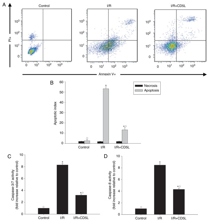
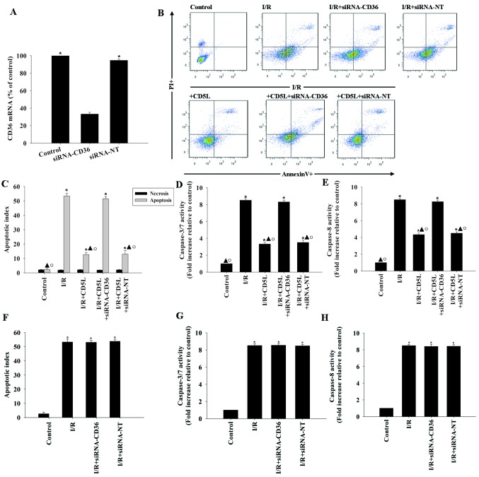
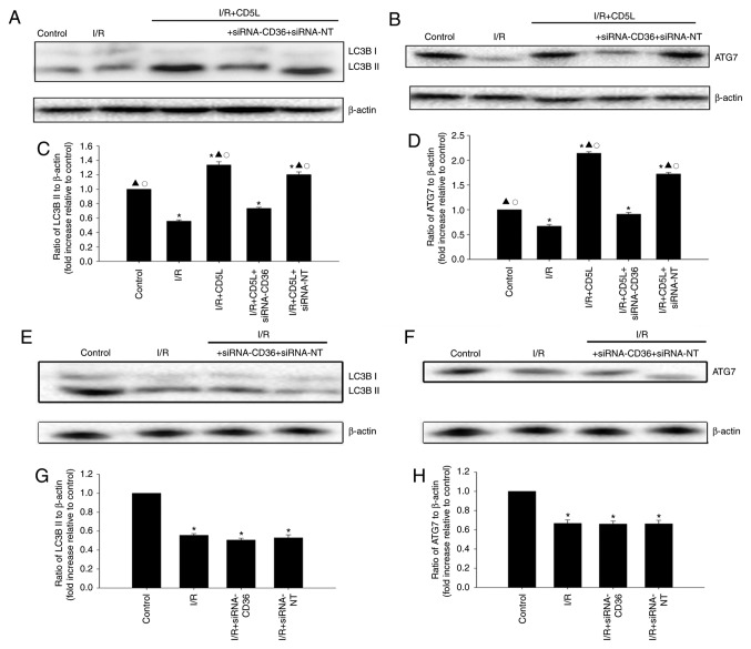
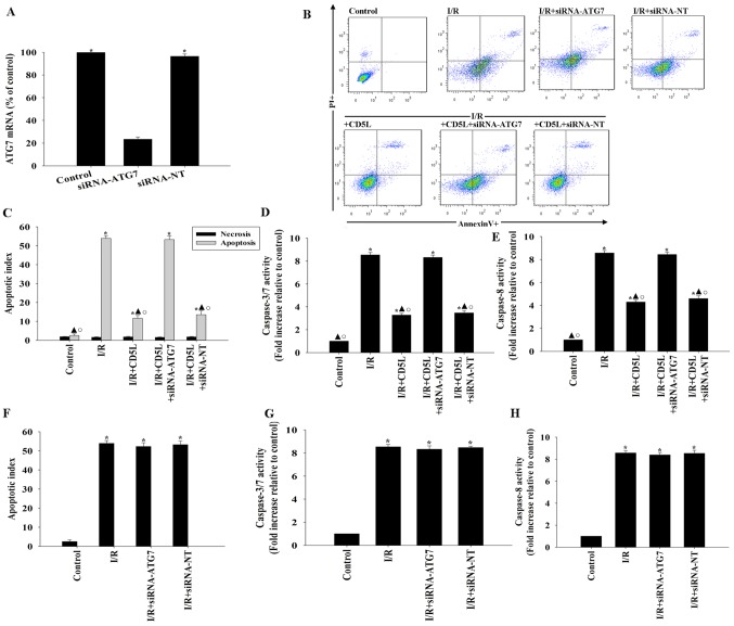
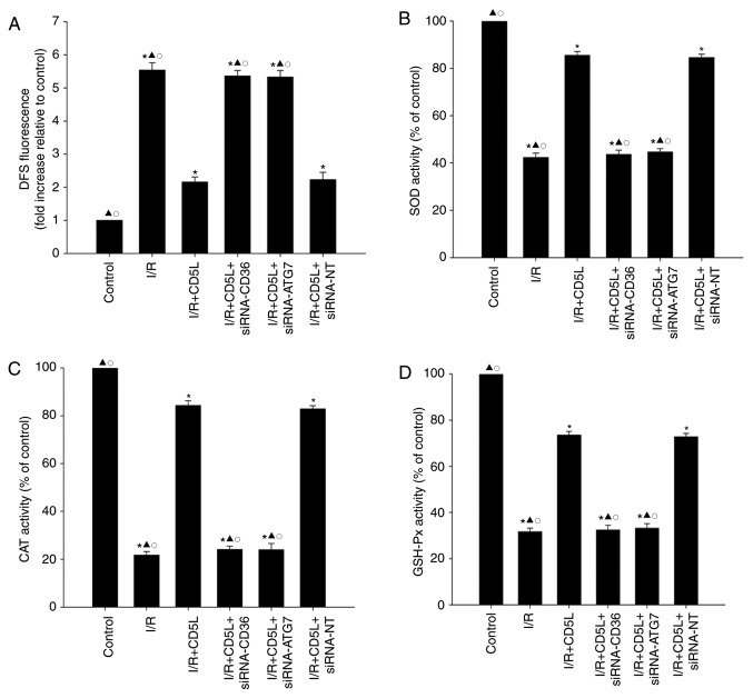
Hepatic ischemia/reperfusion (I/R) injury is a side effect of major liver surgery that is difficult to prevent. I/R injury induces metabolic strain on hepatocytes and limits the tolerable ischemia during liver resection, as well as preservation times during transplantation. Additionally, I/R injury induces apoptosis in hepatocytes. CD5-like (CD5L), an inducer of autophagy, is a soluble scavenger cysteine-rich protein that modulates hepatocyte apoptosis. The aim of the present study was to determine if pharmacologic CD5L was protective against hepatic ischemia-reperfusion injury. Hepatocytes were subjected to I/R culture conditions, and apoptosis and caspase family activity were measured after I/R to model hepatic injury. Treatment with recombinant CD5L significantly suppressed apoptosis and caspase activity through modulating cellular autophagy to maintain activation of the cluster of differentiation 36 (CD36)-dependent autophagy-related 7 (ATG7) signaling pathway. The regulation loop between CD5L and the autophagy signaling pathway was identified to be associated with the inhibition of oxidative stress. Treatment with CD5L significantly inhibited cellular oxidative stress, which was confirmed by silencing the CD36 receptor or the autophagy related protein ATG7 using small interfering RNA, which reversed the antiapoptotic and antioxidative effects of CD5L on hepatocytes under I/R conditions. The results of the present study suggested that CD5L-mediated attenuation of hepatic I/R injury occurs through the CD36-dependent ATG7 pathway, accompanied by the inhibition of oxidative stress, which is associated with enhanced autophagy. In conclusion, the present study identifies CD5L as a novel therapeutic agent for hepatic I/R injury.
Keywords: CD5L; apoptosis; autophagy; hepatic ischemia/reperfusion injury; oxidative stress.
Copyright: © Li et al.
Figures
Similar articles
-
The human CD5L/AIM-CD36 axis: A novel autophagy inducer in macrophages that modulates inflammatory responses.Autophagy. 2015;11(3):487-502. doi: 10.1080/15548627.2015.1017183. Autophagy. 2015. PMID: 25713983 Free PMC article.
-
Long noncoding RNA HOTAIR regulates autophagy via the miR-20b-5p/ATG7 axis in hepatic ischemia/reperfusion injury.Gene. 2019 Feb 20;686:56-62. doi: 10.1016/j.gene.2018.10.059. Epub 2018 Oct 24. Gene. 2019. PMID: 30367982
-
CD5L Promotes M2 Macrophage Polarization through Autophagy-Mediated Upregulation of ID3.Front Immunol. 2018 Mar 12;9:480. doi: 10.3389/fimmu.2018.00480. eCollection 2018. Front Immunol. 2018. PMID: 29593730 Free PMC article.
-
Multifaceted Roles of CD5L in Infectious and Sterile Inflammation.Int J Mol Sci. 2021 Apr 15;22(8):4076. doi: 10.3390/ijms22084076. Int J Mol Sci. 2021. PMID: 33920819 Free PMC article. Review.
-
New Insights Into the Role of Autophagy in Liver Surgery in the Setting of Metabolic Syndrome and Related Diseases.Front Cell Dev Biol. 2021 Jun 1;9:670273. doi: 10.3389/fcell.2021.670273. eCollection 2021. Front Cell Dev Biol. 2021. PMID: 34141709 Free PMC article. Review.
Cited by
-
Potential protective effect of hesperidin on hypoxia/reoxygenation-induced hepatocyte injury.Exp Ther Med. 2021 Jul;22(1):764. doi: 10.3892/etm.2021.10196. Epub 2021 May 13. Exp Ther Med. 2021. PMID: 34035861 Free PMC article.
-
CD36 in liver diseases.Hepatol Commun. 2025 Jan 7;9(1):e0623. doi: 10.1097/HC9.0000000000000623. eCollection 2025 Jan 1. Hepatol Commun. 2025. PMID: 39774047 Free PMC article. Review.
-
Progress in Natural Compounds/siRNA Co-delivery Employing Nanovehicles for Cancer Therapy.ACS Comb Sci. 2020 Dec 14;22(12):669-700. doi: 10.1021/acscombsci.0c00099. Epub 2020 Oct 23. ACS Comb Sci. 2020. PMID: 33095554 Free PMC article. Review.
-
CD5L associates with IgM via the J chain.Nat Commun. 2024 Sep 27;15(1):8397. doi: 10.1038/s41467-024-52175-y. Nat Commun. 2024. PMID: 39333069 Free PMC article.
-
Molecular Regulation Mechanism of Microglial Autophagy in the Pathology of Alzheimer's Disease.Aging Dis. 2023 Aug 1;14(4):1166-1177. doi: 10.14336/AD.2023.0106. Aging Dis. 2023. PMID: 37163443 Free PMC article. Review.
References
-
- Starlinger P, Assinger A, Haegele S, Wanek D, Zikeli S, Schauer D, Birner P, Fleischmann E, Gruenberger B, Brostjan C, Gruenberger T. Evidence for serotonin as a relevant inducer of liver regeneration after liver resection in humans. Hepatology. 2014;60:257–266. doi: 10.1002/hep.26950. - DOI - PubMed
-
- Duval H, Mbatchi SF, Grandadam S, Legendre C, Loyer P, Ribault C, Piquet-Pellorce C, Guguen-Guillouzo C, Boudjema K, Corlu A. Reperfusion stress induced during intermittent selective clamping accelerates rat liver regeneration through JNK pathway. J Hepatol. 2010;52:560–569. doi: 10.1016/j.jhep.2010.01.013. - DOI - PubMed
LinkOut - more resources
Full Text Sources
Miscellaneous