

Prominent Thebesian veins, a rare congenital coronary anomaly presenting as acute myocardial ischemia
- PMID: 32256858
- PMCID: PMC7125364
- DOI: 10.1016/j.jccase.2019.11.007
Prominent Thebesian veins, a rare congenital coronary anomaly presenting as acute myocardial ischemia
Abstract
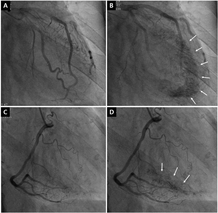
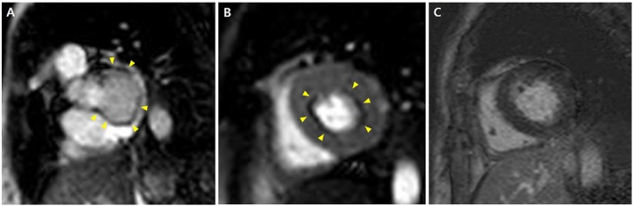
Persistent Thebesian veins with the appearance of multiple coronary artery microfistulas are a rare finding and little is known about their physiologic and clinical features. In addition, few reports have demonstrated the perfusion status of patients with Thebesian veins. We report a 75-year-old woman referred for non-ST-elevation myocardial infarction due to prominent Thebesian veins who displayed a perfusion defect in cardiac magnetic resonance imaging. <Learning objective: This case emphasizes that non-obstructive coronary arterial anomaly can lead to myocardial ischemia with angina symptoms and cardiac enzyme rise. Moreover, it provided the typical angiographic appearance of Thebesian vein network and image of myocardial perfusion defect induced by coronary steal.>.
Keywords: Acute myocardial infarction; Cardiac magnetic resonance imaging; Thebesian veins.
© 2019 Published by Elsevier Ltd on behalf of Japanese College of Cardiology.
Figures

References
-
- Yamanaka O., Hobbs R.E. Coronary artery anomalies in 126,595 patients undergoing coronary arteriography. Cathet Cardiovasc Diagn. 1990;21:28–40. - PubMed
-
- Ansari A. Anatomy and clinical significance of ventricular Thebesian veins. Clin Anat. 2001;14:102–110. - PubMed
-
- Hong G.R., Choi S.H., Kang S.M., Lee M.H., Rim S.J., Jang Y.S. Multiple coronary artery-left ventricular microfistulae in a patient with apical hypertrophic cardiomyopathy: a demonstration by thoracic colar Doppler echocardiography. Yonsei Med J. 2003;44:710–714. - PubMed
-
- Jacob M.A., Goyal S.B., Pacifico L., Spodick D.H. Multiple coronary artery-left ventricular fistulas associated with hereditary hemorrhagic telangiectasia. Chest. 2001;120:1415–1417. - PubMed
Publication types
LinkOut - more resources
Full Text Sources
