. 2020 Mar 24;11(12):1017-1036.
doi: 10.18632/oncotarget.27508.

Roscovitine enhances all- trans retinoic acid (ATRA)-induced nuclear enrichment of an ensemble of activated signaling molecules and augments ATRA-induced myeloid cell differentiation

Affiliations

Roscovitine enhances all- trans retinoic acid (ATRA)-induced nuclear enrichment of an ensemble of activated signaling molecules and augments ATRA-induced myeloid cell differentiation

Asif Rashid et al. Oncotarget. .

Abstract

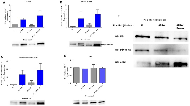
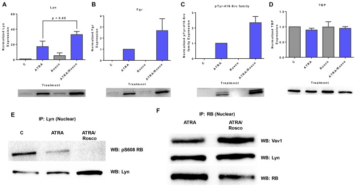
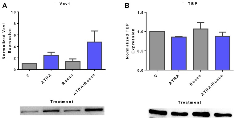
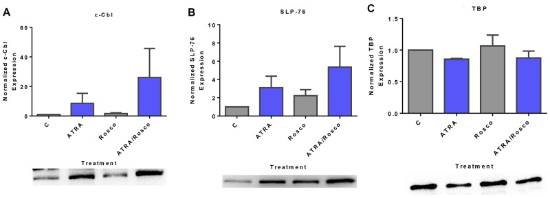
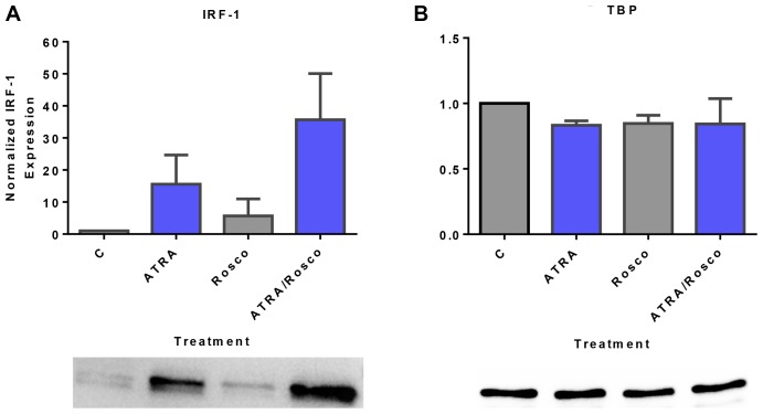
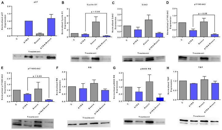
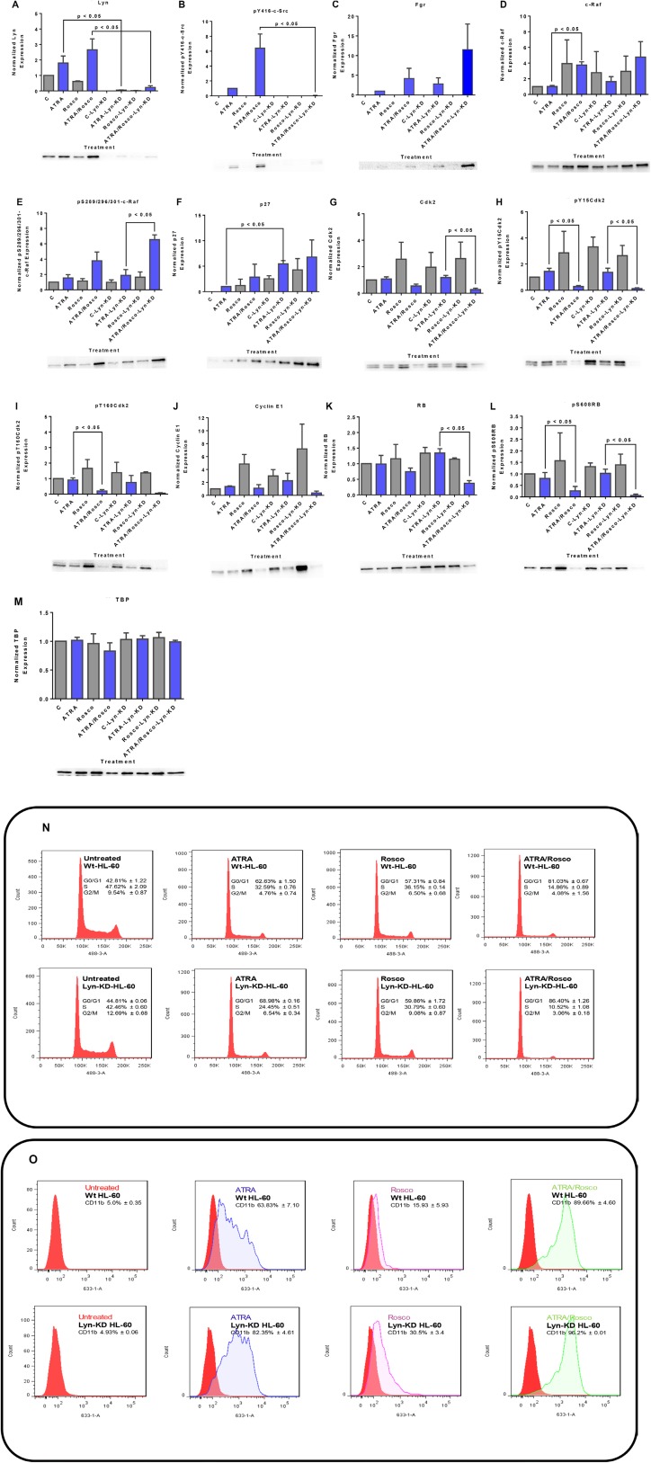
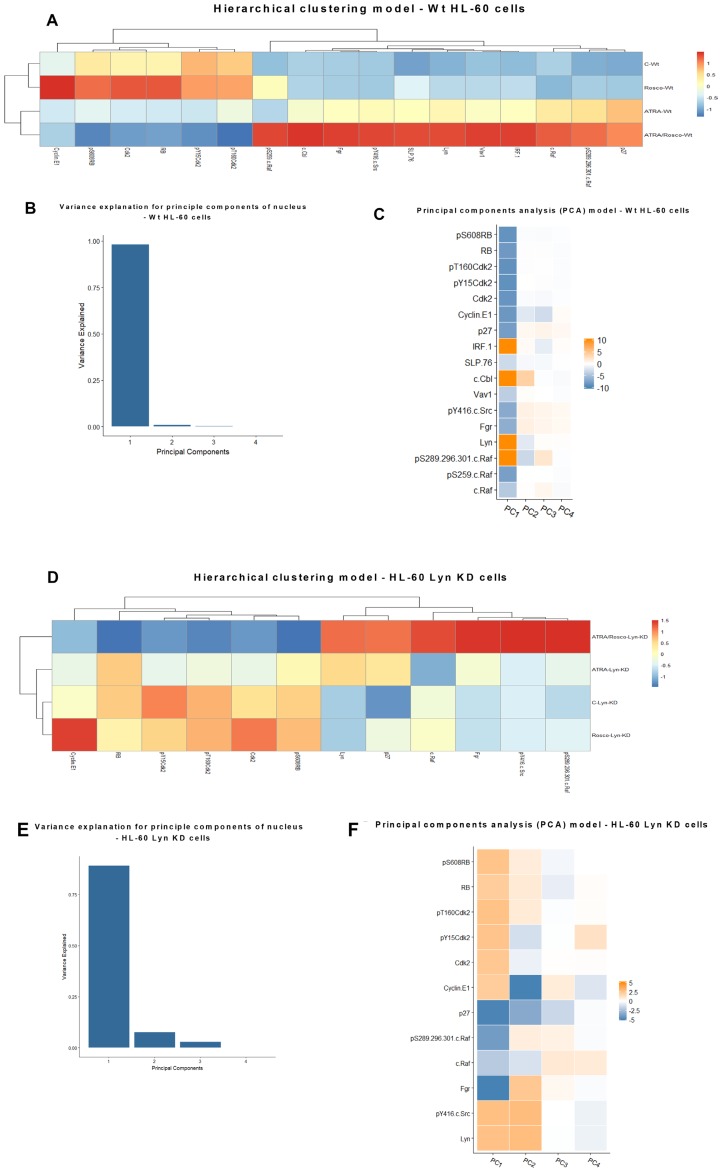
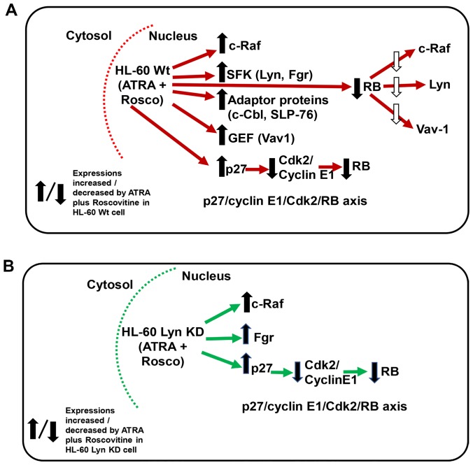
Although ATRA represents a successful differentiation therapy for APL, it is largely ineffective for non-APL AMLs. Hence combination therapies using an agent targeting ATRA-regulated molecules that drive cell differentiation/arrest are of interest. Using the HL-60 human non-APL AML model where ATRA causes nuclear enrichment of c-Raf that drives differentiation/G0-arrest, we now observe that roscovitine enhanced nuclear enrichment of certain traditionally cytoplasmic signaling molecules and enhanced differentiation and cell cycle arrest. Roscovitine upregulated ATRA-induced nuclear c-Raf phosphorylation at S259 and S289/296/301. Nuclear c-Raf interacted with RB protein and specifically with pS608RB, the hinge region phosphorylation controlling E2F binding and cell cycle progression. ATRA-induced loss of pS608RB with cell cycle arrest was associated with loss of RB-sequestered c-Raf, thereby coupling cell cycle arrest and increased availability of c-Raf to promote differentiation. Part of this mechanism reflects promoting cell cycle arrest via ATRA-induced upregulation of the p27 Kip1 CDKI. Roscovitine also enhanced the ATRA-induced nuclear enrichment of other signaling molecules traditionally perceived as cytoplasmic promoters of proliferation, but now known to promote differentiation; in particular: SFKs, Lyn, Fgr; adaptor proteins, c-Cbl, SLP-76; a guanine exchange factor, Vav1; and a transcription factor, IRF-1. Akin to c-Raf, Lyn bound to RB, specifically to pS608RB. Lyn-pS608RB association was greatly diminished by ATRA and essentially lost in ATRA plus roscovitine treated cells. Interestingly Lyn-KD enhanced such ATRA-induced nuclear signaling and differentiation and made roscovitine more effective. ATRA thus mobilized traditionally cytoplasmic signaling molecules to the nucleus where they drove differentiation which were further enhanced by roscovitine.

Keywords: APL; ATRA; HL-60; Lyn; roscovitine.


Conflict of interest statement

CONFLICTS OF INTEREST Authors declare that they have no conflicts of interest with the contents of this article.

Figures

Figure 1
Figure 1. Roscovitine enhances the amount of ATRA-induced phosphorylated c-Raf and phosphorylated c-Raf in the nucleus modulates the RB protein functions.
(AC) Western blot of c-Raf and its phospho-regulatory residues in HL-60 cells cultured with ATRA for 72 h showed that ATRA upregulated nuclear c-Raf, pS259 and pS289/296/301 c-Raf expression and co-treatment with ATRA plus roscovitine further increased of c-Raf and its active phosphorylation sites, pS259 and pS289/296/301, compared to ATRA alone. (D) TATA binding protein (TBP) is the loading control. (E) c-Raf immunoprecipitates probed for RB or S608 RB show that roscovitine enhances ATRA-induced downregulation of the amount of nuclear c-Raf complexed with RB and specifically with its serine 608 phosphorylated form (pS608 RB). An equal amount of pre-cleared nuclear lysate was collected 72 h post treatment and incubated overnight with 2.5 μg of the precipitating antibody with magnetic beads and resolved on 12 % polyacrylamide gels. All blots shown are representative of three replicates.
Figure 2
Figure 2. Roscovitine enhances the expression of ATRA-induced enrichment of nuclear Src-family kinase members.
Nuclear lysates collected after 72 h of treatment were resolved on 12% polyacrylamide gels. 25 μg protein was loaded per well. (A–C) Roscovitine enhances ATRA-induced nuclear Lyn, Fgr and Y416-c-Src expression. p < 0.05 comparing ATRA-treated samples to ATRA/roscovitine-treated samples. (D) TATA binding protein (TBP) was the loading control. (E) Roscovitine augments ATRA-induced reduction of nuclear Lyn interaction with pS608 phosphorylated RB tumor suppressor protein. Co-immunoprecipitation was done using Lyn as bait. (F) Nuclear RB binds Lyn in ATRA and ATRA plus roscovitine treated cells. Co-immunoprecipitation was done in treated cells using RB as bait. Vav also binds RB in these cells. An equal amount of pre-cleared nuclear lysate was collected 72 h post treatment and incubated overnight with 1:100 concentration of the precipitating antibody with magnetic beads and resolved on 12 % polyacrylamide gels. All blots shown are representative of three replicates.
Figure 3
Figure 3. Roscovitine increases ATRA-induced nuclear expression of Vav1.
(A) Western blots of nuclear lysate shows that ATRA enhances the relative expression of nuclear Vav1 compared to untreated cells and ATRA/roscovitine treated HL-60 cells further increases the level of Vav1 compared to ATRA alone at 72 h. (B) TATA binding protein (TBP) was the loading control. All blots shown are representative of three replicates.
Figure 4
Figure 4. Expression of adaptor proteins (c-Cbl, SLP-76) in the nucleus is enhanced by ATRA and roscovitine.
(A, B) Western blots of nuclear lysate show that ATRA enhances the expression of c-Cbl and SLP-76 compared to untreated cells and co-treatment with ATRA and roscovitine further increased their expression compared to ATRA alone at 72 h. (C). (B) TATA binding protein (TBP) was the loading control. All blots shown are representative of three replicates.
Figure 5
Figure 5. Roscovitine enhances ATRA-induced nuclear enrichment of IRF-1.
(A) Western blots of nuclear lysate show that ATRA enhances the relative expression of IRF-1 compared to untreated cells and cells co-treated with ATRA plus roscovitine further increases the level of IRF-1 compared to ATRA alone at 72 h. (B) TATA binding protein (TBP) was the loading control. All blots shown are representative of three replicates.
Figure 6
Figure 6. Roscovitine effects on ATRA-induced changes in nuclear expression of G1/G0 regulatory molecules: p27/cyclin E1/Cdk2/RB pathway.
(A) Western blot of p27Kip1 in cells cultured for 72 h showed that ATRA enhanced nuclear p27Kip1 level, and cells co-treated with ATRA and roscovitine modestly further upregulated the p27Kip1 expression. (B) Roscovitine further decreased ATRA-induced reduction of nuclear cyclin E1 expression in these cells. (C–E) Roscovitine diminishes ATRA-induced downregulation of nuclear CDK2 and specifically its pY15CDK2 and pT160CDK2 phosphorylated forms. (F, G) ATRA plus roscovitine downregulates total RB and pS608 phosphorylated RB. Roscovitine enhanced ATRA-induced reduction of RB phosphorylated at pS608 site. Surprisingly, at the same dose, roscovitine alone enhances nuclear cyclin E1 and CDK2 expression. p < 0.05 comparing ATRA-treated samples to ATRA/Roscovitine-treated samples. (H) TATA binding protein (TBP) was the loading control; a minor artifact caused during image capture can be seen. All blots shown are representative of three replicates.
Figure 7
Figure 7. Western blots of nuclear lysate, CD11b and DNA histograms show that Lyn knockdown enhances ATRA-roscovitine induced gene expression and myeloid differentiation.
(A, B) ATRA and ATRA plus roscovitine co-treated Lyn KD cells caused a modest increase in Lyn and the phospho-residue, pY416-c-Src, expression. (C) Lyn knockdown enhances Fgr expression in Lyn KD cells co-treated with ATRA and roscovitine. (D, E) Lyn knockdown enhances c-Raf and phospho- S289/296/301-c-Raf expression in Lyn KD cells co-treated with ATRA and Roscovitine (F). p27Kip1 protein expression upregulates in Lyn KD cells co-treated with ATRA and roscovitine (GJ). Total Cdk2, phospho-Cdk2Y15, phospho-Cdk2T160 and cyclin E1 downregulates in Lyn KD cells co-treated with ATRA and roscovitine (K, L). Cotreatment of Lyn KD cells also deceases total RB expression and induced hypophosphorylation at serine 608 (S608) of RB (M). TATA binding protein (TBP) was used as loading control. p < 0.05 comparing ATRA-treated samples to ATRA/Roscovitine-treated samples of either wild-type and Lyn-KD cells. (N) Cell cycle distribution showing the percentage of cells in G1/G0, S and G2/M was analyzed using flow cytometry with propidium iodide. ATRA/roscovitine-induced differentiation of Lyn KD stable cells marked by G1/G0 cell cycle arrest was enhanced compared to parental wild type cells. (O) CD11b expression assessed by flow cytometry with an APC-conjugated antibody. Lyn KD cells show enhanced CD11b expression in response to ATRA when compared to wild-type cells. Also, a difference in CD11b expression was found between ATRA/roscovitine-treated parental wild type and Lyn KD cells. All experiments shown are representative of three replicates..
Figure 8
Figure 8. Hierarchical clustering and principal components analysis (PCA) of nuclear protein expression/activation in HL-60 wt and Lyn KD cells.
(A) Clustering based on the expression and activation of nuclear molecules for HL-60 cells either untreated or treated with ATRA, roscovitine or ATRA plus roscovitine, was performed using the ‘pheatmap’ function available in the R package. (B) Contribution by principal components for HL-60 wt cells. (C) Molecular contributors of principal components and their coupling (coupling shown by color similarity) for HL-60 wt cells. (D) Hierarchical clustering of nuclear protein expression/activation for HL-60 Lyn KD cells. Clustering based on the expression and activation of nuclear molecules for HL-60 Lyn KD cells either untreated or treated with ATRA, roscovitine or ATRA plus roscovitine, was performed using the ‘pheatmap’ function available in the R package. (E) Contribution by principal components for HL-60 Lyn KD cells. (F) Molecular contributors of principal components and their coupling (by color) for HL-60 Lyn KD.
Figure 9
Figure 9
(A and B) Schematic diagram of ATRA-roscovitine induced modulation of nuclear molecules in wild-type and Lyn-KD HL-60 cells. Red/Green arrow shows flow of either Wt or Lyn KD HL-60 cells. Up/down arrows show effects shown to be affected by adding ATRA plus Roscovitine.

Similar articles

Cited by

References

    1. Distel E, Cadoudal T, Collinet M, Park EA, Benelli C, Bortoli S. Early induction of pyruvate dehydrogenase kinase 4 by retinoic acids in adipocytes. Mol Nutr Food Res. 2017; 61:1–9. 10.1002/mnfr.201600920. - DOI - PubMed
    1. Ross SA, Mccaffery PJ, Drager UC, De Luca LM. Retinoids in embryonal development. Physiol Rev. 2000; 80:1021–54. 10.1152/physrev.2000.80.3.1021. - DOI - PubMed
    1. Huang ME, Ye YC, Chen SR, Chai JR, Lu JX, Zhoa L, Gu LJ, Wang ZY. Use of all-trans retinoic acid in the treatment of acute promyelocytic leukemia. Blood. 1988; 72:567–72. 10.1182/blood.V72.2.567.567. - DOI - PubMed
    1. Lo-Coco F, Avvisati G, Vignetti M, Thiede C, Orlando SM, Iacobelli S, Ferrara F, Fazi P, Cicconi L, Di Bona E, Specchia G, Sica S, Divona M, et al. , and Gruppo Italiano Malattie Ematologiche dell’Adulto, and German-Austrian Acute Myeloid Leukemia Study Group, and Study Alliance Leukemia . Retinoic acid and arsenic trioxide for acute promyelocytic leukemia. N Engl J Med. 2013; 369:111–21. 10.1056/NEJMoa1300874. - DOI - PubMed
    1. Grignani F, Fagioli M, Alcalay M, Longo L, Pandolfi PP, Donti E, Biondi A, Lo Coco F, Grignani F, Pelicci PG. Acute promyelocytic leukemia: from genetics to treatment. Blood. 1994; 83:10–25. 10.1182/blood.V83.1.10.10. - DOI - PubMed