. 2020 Mar 24;11(12):1051-1074.
doi: 10.18632/oncotarget.27511.

hPCL3S promotes proliferation and migration of androgen-independent prostate cancer cells

Affiliations

hPCL3S promotes proliferation and migration of androgen-independent prostate cancer cells

Souhila Abdelfettah et al. Oncotarget. .

Abstract

Polycomb repressive complex 2 (PRC2) allows the deposition of H3K27me3. PRC2 facultative subunits modulate its activity and recruitment such as hPCL3/PHF19, a human ortholog of Drosophila Polycomb-like protein (PCL). These proteins contain a TUDOR domain binding H3K36me3, two PHD domains and a "Winged-helix" domain involved in GC-rich DNA binding. The human PCL3 locus encodes the full-length hPCL3L protein and a shorter isoform, hPCL3S containing the TUDOR and PHD1 domains only. In this study, we demonstrated by RT-qPCR analyses of 25 prostate tumors that hPCL3S is frequently up-regulated. In addition, hPCL3S is overexpressed in the androgen-independent DU145 and PC3 cells, but not in the androgen-dependent LNCaP cells. hPCL3S knockdown decreased the proliferation and migration of DU145 and PC3 whereas its forced expression into LNCaP increased these properties. A mutant hPCL3S unable to bind H3K36me3 (TUDOR-W50A) increased proliferation and migration of LNCaP similarly to wt hPCL3S whereas inactivation of its PHD1 domain decreased proliferation. These effects partially relied on the up-regulation of genes known to be important for the proliferation and/or migration of prostate cancer cells such as S100A16, PlexinA2, and Spondin1. Collectively, our results suggest hPCL3S as a new potential therapeutic target in castration resistant prostate cancers.

Keywords: PHF19; PRC2; hPCL3S; prostate cancer; β-catenin.


Conflict of interest statement

CONFLICTS OF INTEREST The authors declare no disclosure of potential conflicts of interest.

Figures

Figure 1
Figure 1. Human hPCL3L and hPCL3S isoforms and their identified functional domains.
(A) Comparative structure of hPCL3L and hPCL3S. The structure of the full length human hPCL3L and of the shortest isoform hPCL3S generated by alternative splicing and Alternative Polyadenylation in the coding region (CR-APA) is schematically drawn [21, 22]. The various functional domains are indicated: the TUDOR domain; the two PHD (Plant Homeo domain); the Winged Helix domain involved in the binding to G-C-rich sequences [19, 20] and initially identified as an extended region of Homology (EH) with the Drosophila PCL protein [21] and finally the Reverse Chromodomain (RC) involved in the competitive interaction with AEBP2 for SUZ12 [15]. Two putative nuclear localization signals identified in hPCL3L are also indicated [21]. (B) Structure of the various hPCL3S constructs used in this study. Schematic structure of the wt hPCL3S-AMTag protein and mutants thereof (ΔC-term, W50A and Mut-PHD1). The C-terminal AM-Tag is shown as an open box.
Figure 2
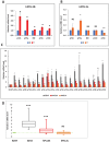
Figure 2. Analyses of hPCL3S and hPCL3L expression in human prostate cancer tissue.
(A–B) Comparison of hPCL3S and hPCL3L mRNA levels in five matched prostate cancer (T) and normal adjacent (N) tissue samples by RT-qPCR analyses. (C) Expression in 20 tumor tissues of hPCL3S, hPCL3L and EZH2. The hPCL3S, hPCL3L and EZH2 expression were measured by RT-qPCR analyses in comparison with their expression in a normal prostate tissue obtained from a young 24-years old donor (BioChain). (D) Quantification of EZH2, hPCL3S and hPCL3L expression in the 20 prostate tumors. The hPCL3S, hPCL3L and EZH2 expression measured in panel C is represented as box plots. The box area corresponds to the first and third quartile. The median is shown as a horizontal line in the box. The maximum and minimum of the values are indicated by the whiskers above and below the box.
Figure 3
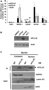
Figure 3. hPCL3S expression is specifically elevated in androgeno-negative prostate cancer cells, in vitro.
(A) The expression levels of hPCL3S, hPCL3L, and EZH2 were examined in normal primary (PrEC) and immortalized (RWPE-1) prostate epithelial cells as well as in transformed androgeno-dependent (LNCaP) and androgeno-independent (DU145 and PC3) prostate cancer cell lines by RT-qPCR analyses. (B) The expression levels of the hPCL3S protein in immortalized (RWPE-1) prostate epithelial cells and in transformed (LNCaP, DU145, and PC3) prostate cancer cell lines were examined using Western blotting. (C) Subcellular localization of endogenous hPCL3S proteins by cell fractionation experiments in DU145. Cytoplasmic and nuclear fractions prepared with the nuclear extraction Kit (Millipore) as previously described [22] were immunoprecipitated with rabbit IgG or anti-hPCL3S antibodies, resolved by SDS-PAGE and immunoblotted with anti-hPCL3S antibodies (top panel) to detect the endogenous hPCL3S proteins. To validate the accuracy of the cell fractionation, samples of each fraction were tested by Western blot with anti-GAPDH, anti-LAMIN A/C, and anti-EZH2 antibodies (bottom panels).
Figure 4
Figure 4. hPCL3S is essential for DU145 and PC3 cell proliferation and migration.
(A) Validation of the hPCL3S siRNA efficiency by RT-qPCR analyses of transfected DU145 and PC3 cells. The expression level of hPCL3S was examined in the DU145 (left panel) and PC3 (right panel) prostate cancer cell lines transfected by the sicontrol RNA or each individual siRNA targeting hPCL3S by RT-qPCR analyses. As control, non-transfected LNCaP or LNCaP transfected by the transfection agent (RNAimax) alone were also tested. (B). Validation of the hPCL3S siRNA efficiency by Western blot analyses of transfected DU145 and PC3 cells. RNAs (used in, A) and proteins were simultaneously prepared from the same transfected cells. Total cell extracts were analyzed by Western blotting to confirm the knockdown of hPCL3S. Actin and α-tubulin were used as a loading control. (C–D) Knockdown of hPCL3S inhibited the cell proliferation of DU145 cells (C) and of PC3 cells (D). The proliferation of non-transfected cells and of cells transfected by RNAiMax alone or in combination with the indicated siRNA was examined using the Incucyte system. (E–F) Overexpression of hPCL3S inhibited the cell migration of DU145 cells (E) and of PC3 cells (E). The migration of non-transfected cells and of cells transfected by RNAiMax alone or in combination with the indicated siRNA was examined with the Incucyte Scratch wound system.
Figure 5
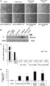
Figure 5. Selection of stable clones expressing the empty vector (p-AM) or the various versions of hPCL3S-AMTag fusion proteins by transfection selected a subpopulation of LNCaP cells.
(A) Transfection of LNCaP with p-AMTag expression vectors selects a cell population with an epithelium-like morphology. Bright field images of parental LNCaP cells, of a pool of clones obtained after transfection of these LNCaP cells by the p-AM empty vector and of individual clones used in this study and obtained after transfection of hPCL3S wt (clones 12 and 17), hPCL3S W50A (clones 5 and 14) and hPCL3S Mut-PHD1 (clones 8 and 20). (B) Imunoblotting analyses of lysates prepared from LNCaP or LNCaP clones obtained after transfection with the various p-AM-Tag expression vectors. Immunoprecipitation analyses were performed with normal rabbit Immunoglobulins (IgG) or with commercial rabbit antibodies generated against anti GST-PCL3S (3S) and followed by Western blot analyses with commercial goat antibodies generated against a C-terminal-peptide (hPCL3S). Please note that this experimental strategy did not allow visualizing the Delta-C-term E mutant. A faint non-specific band is detected in the wtCl2 IgG lane and in the hPCL3S (3S) lanes for empty vector and Delta C-term E. 2% of each total lysates before immunoprecipitation was kept as Input and analyzed by Western blotting with actin antibodies as loading control. (C) RT-qPCR analyses of AR and PSA expression level in the parental LNCaP cells, in LNCaP transfected by the empty vector as well as in the various hPCL3S (wt and mutants) stable clones obtained. (D) Similar RT-qPCR analyses for ALDH1A1 expression level in the indicated cells.
Figure 6
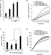
Figure 6. hPCL3S promotes proliferation and migration in the androgeno-dependent human prostate cancer cells, LNCaP.
(A) Stable overexpression of hPCL3S in LNCaP prostate cancer cells. Quantitative PCR analyses of hPCL3S expression was performed on LNCaP cells stably transfected with the empty vector (pool of clones) or on individual clones obtained after transfection of the hPCL3S-AMTag expression vector. (B) Immunofluorescence analyses of LNCaP-hPCL3S-clone 12. The three top panels correspond to the DAPI staining, the conventional immunofluorescence analysis with the anti-AMTag antibody and the merging of the two images, respectively. The bottom panel represents the same experiment except that the primary anti-AMTag antibody was omitted (negative control). (C) Overexpression of hPCL3S promoted the cell proliferation of LNCaP cells. The proliferation of parental LNCaP cells as well of a pool of empty vector transfected clones or the hPCL3S overexpressing clones 12 and 17 was examined using the Incucyte system. (D) Clonogenicity assays. The empty vector transfected cells and the hPCL3S overexpressing clones 12 and 17 were compared in a clonogenicity assay. An example of the crystal blue staining (1 picture out of three) is shown as well as a graphical view of the three samples for each condition (see Supplementary Figure 3 for details). (E) Overexpression of hPCL3S promoted the cell proliferation of LNCaP cells in hormone-depleted medium. The proliferation of parental LNCaP cells as well of a pool of empty vector transfected clones or the hPCL3S overexpressing clones 12 grown in normal medium (RPMI 1640 +10% fetal calf serum) or in hormone-depleted medium (RPMI 1640 +10% charcoal-stripped fetal calf serum) was examined using the Incucyte system. (F) Overexpression of hPCL3S promoted the cell migration of LNCaP cells. The migration of parental LNCaP cells as well of the various stable clones was examined using the Incucyte Scratch Wound system.
Figure 7
Figure 7. The short (AA 155-207) specific C-terminal end of hPCL3S generated by the alternative polyadenylation mechanism is not essential for the promotion of proliferation and migration.
(A) Stable overexpression of hPCL3S-Delta C-term in two individual clones was analyzed by RT-qPCR analyses. (B) Overexpression of hPCL3S-ΔC-Term promoted the proliferation of LNCaP cells as efficiently as wt PCL3S. The proliferation of LNCaP transfected by the empty vector, of hPCL3S overexpressing clone12 and of two clones overexpressing the Delta C-term mutant was examined using the Incucyte system. (C) The effect of the overexpression of hPCL3S-ΔC-Term was examined using the clonogenicity assay (the original pictures used for this graphical view are shown in Supplementary Figure 3). (D) Overexpression of hPCL3S-ΔC-Term promoted the cell migration of LNCaP cells as efficiently as wt hPCL3S. The migration of the various indicated clones was examined using the Incucyte Scratch Wound system.
Figure 8
Figure 8. The H3K36me3 binding activity of the hPCL3S TUDOR domain is not required for the promotion of proliferation and migration.
(A) Stable overexpression of hPCL3S-W50A in two individual clones was confirmed by RT-qPCR analyses. (B) The W50A point mutation in the TUDOR domain did not impaired the proliferation of LNCaP cells as compared with wt hPCL3S. The proliferation of empty vector transfected cells or wt hPCL3S overexpressing clone12 was compared to two different clones overexpressing hPCL3S-W50A (clones 5 and 14) using the Incucyte system. (C) The effect of the overexpression of hPCL3S W50A was examined using the clonogenicity assay (the original pictures used for this graphical view are shown in Supplementary Figure 4). (D) Overexpression of hPCL3S W50A promoted the cell migration of LNCap cells as efficiently as wt PCL3S. The migration of the various stable clones was examined using the Incucyte Scratch Wound system.
Figure 9
Figure 9. The PHD1 domain is essential for the promotion of proliferation.
(A) Stable overexpression of hPCL3S-PHD1-Mut in two individual clones was confirmed by RT-qPCR analyses. (B) The mutation of an essential β strand in the PHD1 domain impaired the proliferation of LNCaP cells as compared with wt hPCL3S. The proliferation of empty vector transfected cells or wt hPCL3S overexpressing clone12 was compared to two different clones overexpressing hPCL3S-PHD1-Mut (clones 8 and 20) using the Incucyte system. (C) The effect of the overexpression of hPCL3S-PHD1-Mut was examined using the clonogenicity assay (the original pictures used for this graphical view are shown in Supplementary Figure 4). Error bars correspond to standard deviation between the empty vector and each clone. The *** above the brackets corresponds to the comparison of the wt clone 12 with the two PHD1-mutated clones. (D) Overexpression of hPCL3S-PHD1-Mut slightly promoted the cell migration of LNCaP cells as compared to the control empty vector but less efficiently than wt PCL3S. The migration of the various stable clones was examined using the Incucyte Scratch Wound system.
Figure 10
Figure 10. IL6 appeared as a hPCL3S target gene but independently of βcatenin stabilization in DU145 cells.
After transfection of DU145 cells by hPCL3S siRNAs, RNAs and proteins extracts were simultaneously prepared from the same cells. (A) Efficient knockdown of hPCL3S in DU145 after transfection of the three individual siRNAs was confirmed by RT-qPCR analyses. (B) IL6 expression is down regulated after transfection of two out of three individual hPCL3S siRNAs. The RT samples analyzed in panel A were checked for expression of IL6 by qPCR. (C) Efficient knockdown of hPCL3S did not affect the βcatenin protein levels. Total proteins extracts were tested by Western blot for the expression of hPCL3S, βcatenin, and Tubulin as a loading control.
Figure 11
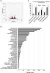
Figure 11. Global analyses of the RNA-Seq data of LNCaP empty vector and LNCaP cells overexpressing hPCL3S (Clone 12).
(A) Volcano plot shows gene expression changes upon PHF19S induction in LNCap cells. X-axis shows fold changes in expression and y-axis shows adjusted p values. Red dots indicate the genes differentially expressed (at least two-fold changes and p-value < 0.05). (B) The enriched pathways were identified using the Database for Annotation, Visualization and Integrated Discovery (DAVID). (C) Graphical representation of the common functions between up- and down-regulated genes.
Figure 12
Figure 12. Validation of some differentially expressed genes.
(A) FAM1484A is up-regulated in LNCaP transfected by the p-AM empty vector but down-regulated in LNCaP clone 12 cells surexpressing hPCL3S. RNAs were extracted from the non-transfected LNCaP cells as well as from cells transfected with the empty vector or with the p-AM-hPCL3S wt clone 12. The expression of Neuron-specific enolase, a classical marker of NED, has also been tested. (B) Validation by RT-qPCR analyses of the RNA-Seq data showing differential regulation of some selected genes in the LNCaP clone 12 cells. Selected genes (S100A16, SPON1 and PLXNA2) were analyzed by RT-qPCR and showed the expected differential regulation.

Similar articles

Cited by

References

    1. Wang G, Zhao D, Spring DJ, DePinho RA. Genetics and biology of prostate cancer. Genes Dev. 2018; 32:1105–1140. 10.1101/gad.315739.118. - DOI - PMC - PubMed
    1. Terry S, Beltran H. The many faces of neuroendocrine differentiation in prostate cancer progression. Front Oncol. 2014; 4:60. 10.3389/fonc.2014.00060. - DOI - PMC - PubMed
    1. Tai S, Sun Y, Squires JM, Zhang H, Oh WK, Liang CZ, Huang J. PC3 is a cell line characteristic of prostatic small cell carcinoma. Prostate. 2011; 71:1668–1679. 10.1002/pros.21383. - DOI - PMC - PubMed
    1. Uysal-Onganer P, Kawano Y, Caro M, Walker MM, Diez S, Darrington RS, Waxman J, Kypta RM. Wnt-11 promotes neuroendocrine-like differentiation, survival and migration of prostate cancer cells. Mol Cancer. 2010; 9:55. 10.1186/1476-4598-9-55. - DOI - PMC - PubMed
    1. Moparthi L, Pizzolato G, Koch S. Wnt activator FOXB2 drives the neuroendocrine differentiation of prostate cancer. Proc Natl Acad Sci U S A. 2019; 116:22189–22195. 10.1073/pnas.1906484116. - DOI - PMC - PubMed