. 2020 Mar 19:18:696-704.
doi: 10.1016/j.csbj.2020.03.009. eCollection 2020.

Crystal structure of the C-terminal domain of DENR

Affiliations

Crystal structure of the C-terminal domain of DENR

Ivan B Lomakin et al. Comput Struct Biotechnol J. .

Abstract

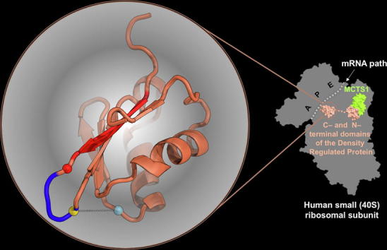
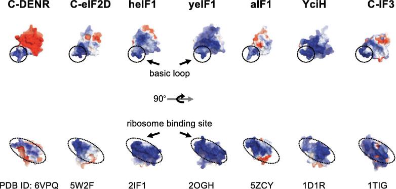
The density regulated protein (DENR) forms a stable heterodimer with malignant T-cell-amplified sequence 1 (MCT-1). DENR-MCT-1 heterodimer then participates in regulation of non-canonical translation initiation and ribosomal recycling. The N-terminal domain of DENR interacts with MCT-1 and carries a classical tetrahedral zinc ion-binding site, which is crucial for the dimerization. DENR-MCT-1 binds the small (40S) ribosomal subunit through interactions between MCT-1 and helix h24 of the 18S rRNA, and through interactions between the C-terminal domain of DENR and helix h44 of the 18S rRNA. This later interaction occurs in the vicinity of the P site that is also the binding site for canonical translation initiation factor eIF1, which plays the key role in initiation codon selection and scanning. Sequence homology modeling and a low-resolution crystal structure of the DENR-MCT-1 complex with the human 40S subunit suggests that the C-terminal domain of DENR and eIF1 adopt a similar fold. Here we present the crystal structure of the C-terminal domain of DENR determined at 1.74 Å resolution, which confirms its resemblance to eIF1 and advances our understanding of the mechanism by which DENR-MCT-1 regulates non-canonical translation initiation and ribosomal recycling.

Keywords: Density regulated protein (DENR); Protein synthesis regulation; Translation initiation; Translation recycling; Translation reinitiation.


Figures

None
Graphical abstract
Fig. 1
Fig. 1
The structure of the C-terminal domain of human DENR110–195. A. Domain organization of human eIF1, eIF2D, MCT-1, and DENR (numbers showing amino acid residues at the borders). Domain swapping and packing of the C-DENR molecules in the: B. P6422 and C. P21 crystal form. D. Superposition of C-DENR molecules: from both crystal forms (left), with the β strand 1 swapped (red), present (middle), with the modeled basic loop (red, right). β strands 1 and 5, and positions of some amino acid residues are marked. (For interpretation of the references to colour in this figure legend, the reader is referred to the web version of this article.)
Fig. 2
Fig. 2
Superposition of the C-DENR110–195 structure (coral) with: A. the structure of the human C-DENR (light gray) bound to the human 40S subunit; B. the NMR structure of the human eIF1 (green, PDB: 2IF1, amino acid residues 28–108); C. the structure of the human C-eIF2D (gray, PDB: 5W2F); D. structures of archaeal aIF1 (blue, PDB: 5ZCY) and bacterial YciH (yellow, PDB: 1D1R). The basic loop and β-hairpin 2 loop are marked by an arrow; N- and C-terminus of a protein are marked by N and C, respectively. Positions of amino acid residues of C-DENR are presented by spheres: Pro121 (red), Lys125 (gold), Cys154 (blue). A crosslink between residues Lys125 and Cys154 is marked by a dotted line. (For interpretation of the references to colour in this figure legend, the reader is referred to the web version of this article.)
Fig. 3
Fig. 3
Sequence alignment of the eIF1-like proteins. Multiple sequence alignment using program Clustal Omega (www.clustal.org/omega) with default settings was performed for protein sequences (accession/organism) of DENR: h (NP_003668.2, Homo sapiens), r (NP_001108518.1, Rattus norvegicus), m (NP_080879.1, Mus musculus), s (NP_001134755.1, Salmo salar), x (NP_001086186.1, Xenopus laevis), d (NP_573176.1, Drosophila melanogaster), y (P47089.1, Saccharomyces cerevisiae); eIF2D: h (P41214.3), y (DAA11962.1); eIF1: h (P41567.1), m (P48024.2), x (NP_001079402.1), d (NP_001079402.1), y (NP_014155.1), t (DAA33997.1, Tetrahymena thermophila); aIF1: m (PDBID 4MO0, Methanocaldococcus jannaschii), p (PDBID 5ZCY, Pyrococcus horikoshii); YciH (VWQ02336.1, Escherichia coli). Conserved amino acid residues are marked in red, amino acid residues involved in rRNA binding are marked by arrows, regions are marked by lines: blue (basic loop), gray (β-hairpin 2 loop), brown (helix α1). (For interpretation of the references to colour in this figure legend, the reader is referred to the web version of this article.)
Fig. 4
Fig. 4
Electrostatic charge distribution of the human C-DENR110–195, C-eIF2 (PDB: 5W2F) and eIF1 (heIF1, PDB: 2IF1), Saccharomyces cerevisiae eIF1 (yeIF1, PDB: 2OGH), Pyrococcus horikoshii aIF1 (PDB: 5ZCY), Escherichia coli YciH (PDB: 1D1R) and Geobacillus stearothermophilus C-IF3 (PDB: 5ZCY). Surfaces are colored by potential on solvent accessible surface.
Fig. 5
Fig. 5
Size-exclusion chromatography analysis and thermal unfolding of the C-DENR mutants using Circular Dichroism. A. Chromatogram of C-DENR mutants runs on the Superdex 75 10/300 column. B. Temperature-dependent CD spectra of C-DENR110–195 (top), C-DENR109–198 (middle), C-DENR106–199 (bottom). C. Temperature-dependent transitions from folded to unfolded state monitored at 220 nm.
Fig. 6
Fig. 6
Free energy landscape of the C-DENR semi-extended conformation. S denotes the starting crystal structure, I-1 and I-2 are the intermediate states and G is the compact ground state. The two collective variables are: β-sheet, β-strand character of the N-terminal residue stretch 121–133 of the C-DENR110–195 mutant; d-COM, the distance of the center of mass of these residues with the rest of the molecule.
Fig. 7
Fig. 7
The electron density map 2Fo-Fc contoured at σ = 1.5 (blue mesh) calculated for the region of C-DENR were the crosslink between Lys125 and oxidized Cys154 (S-hydrohycysteine, Cso154) was observed in the P21 crystal form. (For interpretation of the references to colour in this figure legend, the reader is referred to the web version of this article.)

Similar articles

Cited by

References

    1. Deyo J.E., Chiao P.J., Tainsky M.A. drp, a novel protein expressed at high cell density but not during growth arrest. DNA Cell Biol. 1998;17(5):437–447. - PubMed
    1. Oh J.J. Identification of differentially expressed genes associated with HER-2/neu overexpression in human breast cancer cells. Nucleic Acids Res. 1999;27(20):4008–4017. - PMC - PubMed
    1. Lomakin I.B. Crystal structure of the human ribosome in complex with DENR-MCT-1. Cell Rep. 2017;20(3):521–528. - PMC - PubMed
    1. Skabkin M.A. Activities of Ligatin and MCT-1/DENR in eukaryotic translation initiation and ribosomal recycling. Genes Dev. 2010;24(16):1787–1801. - PMC - PubMed
    1. Weisser, M., et al., Structural and functional insights into human re-initiation complexes. Mol Cell, 2017;67(3): p. 447–456 e7. - PubMed

LinkOut - more resources