

Case Reports
doi: 10.1093/jscr/rjaa037.
eCollection 2020 Mar.
Primary mesenchymal chondrosarcoma of the orbit in a young female: imaging and histopathological features
Affiliations
- PMID: 32257101
- PMCID: PMC7103418
- DOI: 10.1093/jscr/rjaa037
Item in Clipboard
Case Reports
Primary mesenchymal chondrosarcoma of the orbit in a young female: imaging and histopathological features
J Surg Case Rep.
.
Abstract
Mesenchymal chondrosarcoma (MCS) is a rare high-grade sarcoma of bone and soft tissue with highly aggressive behavior and a peak incidence in the second and third decades. We report a case of primary orbital MCS in a 30 year-old female, with radiological and clinicopathological features. Orbital MCS is an entity that should be considered in the differential diagnosis of calcified orbital lesions.
Keywords: chondrosarcoma; mesenchymal; orbit.
Published by Oxford University Press and JSCR Publishing Ltd. All rights reserved. © The Author(s) 2020.
Figures
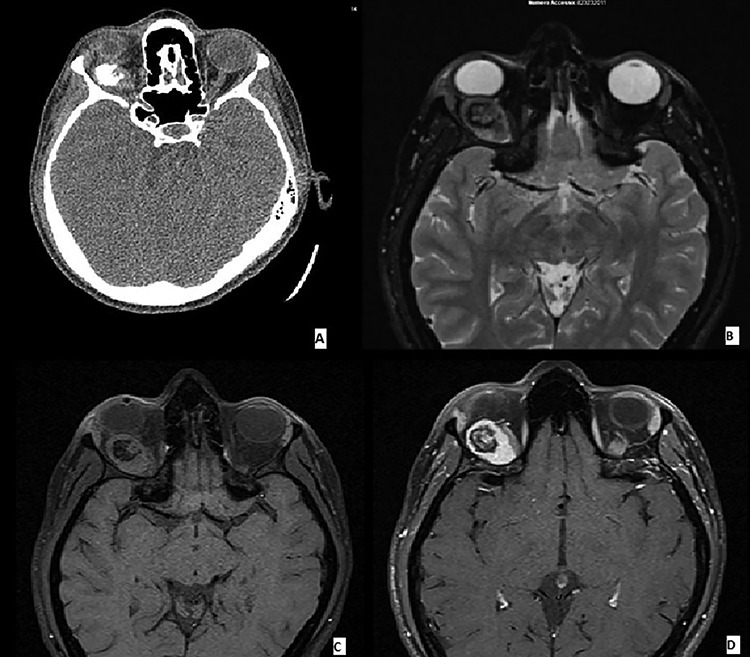
(A–D) Imaging features of MCS: CT scans demonstrated an ovoidal soft tissue mass with central calcification (A), T2 (B) and T1 (C) weighted images showing an oval tumor with iso-hyperintense signal with a calcified central component. Enhanced T1 weighted image demonstrating an omogenous tumoral enhancement except for the central calcified part (D).

Low-power view of the lesion showing central ossification (H&E).

Abrupt transition from small cell component to well differentiated cartilaginous area (H&E, high power view).

Typical hemangiopericytoma-like vascular pattern (H&E, high power view).

Immunohistochemical features of MCS: cytoplasmic positivity for CD99 in the small cell component (IHC stain).

Nuclear positivity for S100 in the cartilaginous component (IHC stain).
References
-
- Lightenstein L, Bernstein D. Unusual benign and malignant chondroid tumors of bone. A survey of some mesenchymal cartilage tumors and malignant chondroblastic tumors, including a few multicentric ones, as well as many atypical benign chondroblastomas and chondromyxoid fibromas. Cancer 1959;12:1142–57. - PubMed
-
- Tos D, Paolo A. Soft Tissue Sarcoma: A Pattern Approach to Diagnosis. Cambridge University Press, 2019, 336–40ISBN 9781107040809
-
- Yang BT, Wang YZ, Wang XY, Wang ZC. Mesenchymal chondrosarcoma of the orbit: CT and MRI findings. Clin Radiol 2012;67:346–51. - PubMed
Publication types
LinkOut - more resources
Full Text Sources
