. 2020 Feb 12;7(2):192054.
doi: 10.1098/rsos.192054. eCollection 2020 Feb.

Control of synchronization ratios in clock/cell cycle coupling by growth factors and glucocorticoids

Affiliations

Control of synchronization ratios in clock/cell cycle coupling by growth factors and glucocorticoids

S Almeida et al. R Soc Open Sci. .

Abstract

The cell cycle and the circadian clock are essential cyclic cellular processes often synchronous in healthy cells. In this work, we use previously developed mathematical models of the mammalian cell cycle and circadian cellular clock in order to investigate their dynamical interactions. Firstly, we study unidirectional cell cycle → clock coupling by proposing a mechanism of mitosis promoting factor (MPF)-controlled REV-ERBα degradation. Secondly, we analyse a bidirectional coupling configuration, where we add the CLOCK : BMAL1-mediated MPF repression via the WEE1 kinase to the first system. Our simulations reproduce ratios of clock to cell cycle period in agreement with experimental observations and give predictions of the system's synchronization state response to a variety of control parameters. Specifically, growth factors accelerate the coupled oscillators and dexamethasone (Dex) drives the system from a 1 : 1 to a 3 : 2 synchronization state. Furthermore, simulations of a Dex pulse reveal that certain time regions of pulse application drive the system from 1 : 1 to 3 : 2 synchronization while others have no effect, revealing the existence of a responsive and an irresponsive system's phase, a result we contextualize with observations on the segregation of Dex-treated cells into two populations.

Keywords: cell cycle; circadian clock; control parameters; coupled oscillators; modelling and simulation; synchronization ratios.


Conflict of interest statement

We declare we have no competing interests.

Figures

Figure 1.
Figure 1.
Schematic of the unidirectional cell cycle → clock coupling mechanism. MPF in its active form represses REV via phosphorylation leading to REV’s subsequent degradation.
Figure 2.
Figure 2.
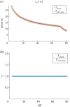
Strong coupling of the circadian clock and cell cycle models by MPF-induced degradation of REV. For cm = 0.2, the system is strongly coupled with 1 : 1 period-lock. The period of the clock follows that of the cell cycle that decreases with GF. Cell cycle oscillations occur for 4 ≤ GF ≤ 80.
Figure 3.
Figure 3.
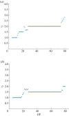
Weak coupling of the circadian clock and cell cycle models by MPF-induced degradation of REV. For (a) cm = 0.04 and (b) cm = 0.08, the system is in weak/moderate coupling and distinct period-lock ratios are obtained depending on GF, forming a pattern similar to that of the devil’s staircase, where the period-lock ratio is increasing but remains constant by intervals of GF. GF and cm are control parameters for the PL ratios. The 3 : 2 experimentally observed PL state is obtained.
Figure 4.
Figure 4.
cm is a control parameter for the period-lock dynamics of the coupled system. Varying cm with fixed GF = 40 causes the ratio of clock to cell cycle period to vary in steps, where the 2 : 1, 3 : 2 and 1 : 1 period-lock ratios are obtained.
Figure 5.
Figure 5.
An input of Dex drives the system from 1 : 1 to 3 : 2 period-lock. With cm = 0.1 and Dex = 0 the system is in strong coupling with 1 : 1 PL for 4 ≤ GF ≤ 24. With cm = 0.1 and Dex = 10 the 3 : 2 PL ratio is obtained.
Figure 6.
Figure 6.
The input IB drives the system from 2 : 1 to 1 : 1 period-lock. With cm = 0.04 and IB = 0 the system period-locks in 2 : 1 for 30 ≤ GF ≤ 60 (see also figure 3). With cm = 0.04 and IB = 10 the 1 : 1 period-lock is obtained.
Figure 7.
Figure 7.
Schematic of the bidirectional coupling mechanism. Bidirectional coupling between the cell cycle and clock oscillators that includes two forms of coupling: MPF phosphorylates REV inducing its degradation and BMAL1 represses MPF by promoting wee1.
Figure 8.
Figure 8.
Period response of the bidirectional coupled system. Our bidirectional coupling mechanisms are able to reproduce the overall observed oscillators’ period response of acceleration with GF and result in a good fit to experimental data [11,18], taking GF=%FBS, with cm = 0.2 and cb = 30.
Figure 9.
Figure 9.
Period-lock for different values of cb and cm with GF = 20. Varying cb and cm for fixed GF = 20 results in different period-lock ratios. In the white region, there is no oscillation. The 1 : 1, 3 : 2, 2 : 1 and 4 : 3 ratios are the most prevalent. An Arnold Tongue pattern is visible for 3 : 2 synchronization.
Figure 10.
Figure 10.
A Dex input induces the system from a 1 : 1 to a 3 : 2 period-lock in bidirectional coupling. With cm = 0.2 and cb = 40 the system locks in 1 : 1 synchronization for 8 ≤ GF ≤ 20. Adding Dex = 5 shifts the system to a 3 : 2 period-lock ratio for the same values of GF.
Figure 11.
Figure 11.
Response curves of the synchronization ratio of the bidirectional coupled system over two periods. A 1 h Dex pulse is applied over the course of two periods. Parameters are cm = 0.2 and cb = 40, GF = 15 and Dexpulse = 40. The system’s synchronization state changes only for certain times of pulse application Tpulse. The responsive phase corresponds to that of increasing BMAL1.
Figure 12.
Figure 12.
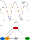
The reduced model can recover the main properties of the circadian clock. (a) Output of the reduced model, oscillations have a period of 18.6 h with parameters of table 2; BMAL1 and PER : CRY maintain an antiphasic oscillation. (b) A scheme of the reduced model. Red dashed arrows show the effect of Eboxes on REV and PER : CRY and can be removed.

References

    1. Takahashi JS. 2017. Transcriptional architecture of the mammalian circadian clock. Nat. Rev. Genet. 18, 164–179. (10.1038/nrg.2016.150) - DOI - PMC - PubMed
    1. Chaix A, Zarrinpar A, Panda S. 2016. The circadian coordination of cell biology. J. Cell Biol. 215, 15–25. (10.1083/jcb.201603076) - DOI - PMC - PubMed
    1. Feillet C, van der Horst GTJ, Levi F, Rand DA, Delaunay F. 2015. Coupling between the circadian clock and cell cycle oscillators: implication for healthy cells and malignant growth. Front. Neurol. 6, 96 (10.3389/fneur.2015.00096) - DOI - PMC - PubMed
    1. Shostak A. 2017. Circadian clock, cell division, and cancer: from molecules to organism. Int. J. Mol. Sci. 18, 873 (10.3390/ijms18040873) - DOI - PMC - PubMed
    1. Matsuo T, Yamaguchi S, Mitsui S, Emi A, Shimoda F, Okamura H. 2003. Control mechanism of the circadian clock for timing of cell division in vivo. Science 302, 255–259. (10.1126/science.1086271) - DOI - PubMed

LinkOut - more resources