Review
. 2020 Feb 26;6(4):FSO465.
doi: 10.2144/fsoa-2019-0116.

Role of exosomes and exosomal microRNAs in cancer

Affiliations
Review

Role of exosomes and exosomal microRNAs in cancer

Nihat Dilsiz. Future Sci OA. .

Abstract

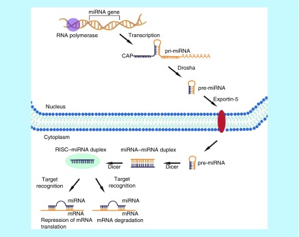
A growing body of evidence indicates that exosomes play a critical role in the cell-cell communication process. Exosomes are biological nanoparticles with an average diameter of 30-100 nm in size and are produced by almost all cell types in the human body; however, cancer cells contain higher concentrations of exosomes than healthy cells. They are released into all body fluids and contain double-stranded DNA (originated from nucleus and mitochondria), a variety of RNA species, and specific protein biomarkers that can be utilized as cancer biomarkers and therapeutic targets, and lipids. Therefore, the specific exosomes secreted by tumor cells could be used to predict the existence of the presence of a tumor in cancer patients. This review summarizes the role of exosomes in cancer development and their potential utility in the clinic.

Keywords: biomarkers; body fluids; cancer; exosomes; extracellular vesicles; miRNA.


Conflict of interest statement

Financial & competing interests disclosure The author has no relevant affiliations or financial involvement with any organization or entity with a financial interest in or financial conflict with the subject matter or materials discussed in the manuscript. This includes employment, consultancies, honoraria, stock ownership or options, expert testimony, grants or patents received or pending, or royalties. No writing assistance was utilized in the production of this manuscript.

Figures

Figure 1.
Figure 1.. The structure and content of exosome.
Exosomes contain various types of proteins, nucleic acids, lipids and metabolites.
Figure 2.
Figure 2.. The formation and releasing of exosome.
(A) Exosome is derived from early endosome formed from plasma membrane. (B) Early endosome becomes late endosomes. (C) Then forms early multivesicular bodies. (D) Late multivesicular bodies. (E) Late multivesicular bodies can either get degraded by lysosomes or fuse with the membrane to release exosomes.
Figure 3.
Figure 3.. Exosomes biogenesis and sorting into the blood.
Exosome can be either fused with lysosomes for degradation or with plasma membrane thereby releasing exosomes to the extracellular space. Cell-released exosomes then can be taken up by neighboring recipient cells or travel through biological fluids such as blood, urine or saliva.
Figure 4.
Figure 4.. Transfer of exosomal miRNA from donor cell to recipient cells.
(A) miRNAs are sorted to exosome during MVB formation. (B) Exosomes are released into the extracellular space. (C) Exosomal miRNAs can be delivered to recipient cells by endocytosis. (D) Fusion of the exosomes with the plasma membrane using soluble N-ethylmaleimide sensitive fusion into cells primarily uses receptor-mediated endocytosis. (E) Exosomes may also bind to a receptor and activate specific signaling pathways. MVB: Multivesicular body.
Figure 5.
Figure 5.. Primary tumor and angiogenesis.
(A) Cell proliferate to form primary tumor formation (without blood vessels). (B) Tumor mass increase and produce angiogenic factors that stimulate new blood vessel formation from the main blood vessel toward the tumor cells.
Figure 6.
Figure 6.. Overview of the different exosome isolation and purification techniques.
Figure 7.
Figure 7.. The biogenesis of microRNA.
Noncoding miRNAs genes are transcribed in the nucleus into primary miRNAs, which are further processed into premary miRNA and then exported into the cytoplasm where they are finally converted into their matured forms. Mature miRNA then bind to its target mRNA with base pairing, acting as negative regulators of mRNA translation (either mRNA degradation or inhibition of protein expression from mRNA).

References

    1. Bray F, Ferlay J, Soerjomataram I, Siegel RL, Torre LA, Jemal A. Global Cancer Statistics 2018: GLOBOCAN estimates of incidence and mortality worldwide for 36 cancers in 185 countries. CA Cancer J. Clin. 68(6), 394–424 (2018). - PubMed
    1. Markopoulos GS, Roupakia E, Tokamani M. et al. A step-by-step microRNA guide to cancer development and metastasis. Cell. Oncol. 40(4), 303–339 (2017). - PubMed
    1. Liu X, Liu X, Wu Y. et al. MicroRNAs in biofluids are novel tools for bladder cancer screening. Oncotarget 8(19), 32370 (2017). - PMC - PubMed
    1. Li W, Li C, Zhou T. et al. Role of exosomal proteins in cancer diagnosis. Mol. Cancer 16(1), 145 (2017). - PMC - PubMed
    1. Tadimety A, Closson A, Li C, Yi S, Shen T, Zhang JX. Advances in liquid biopsy on-chip for cancer management: technologies, biomarkers, and clinical analysis. Crit. Rev. Cl. Lab. Sci. 55(3), 140–162 (2018). - PMC - PubMed

LinkOut - more resources