. 2020 Feb 29:24:109-120.
doi: 10.1016/j.jare.2020.02.014. eCollection 2020 Jul.

Etoricoxib prevents progression of osteolysis in repeated intra-articular monosodium urate-induced gouty arthritis in rats

Affiliations

Etoricoxib prevents progression of osteolysis in repeated intra-articular monosodium urate-induced gouty arthritis in rats

Yen-You Lin et al. J Adv Res. .

Abstract

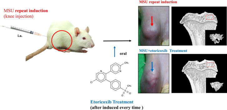
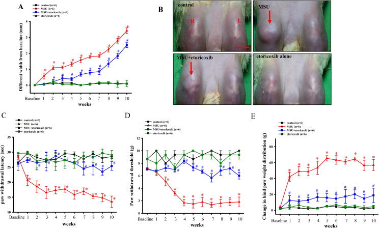
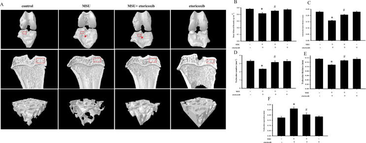
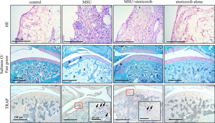
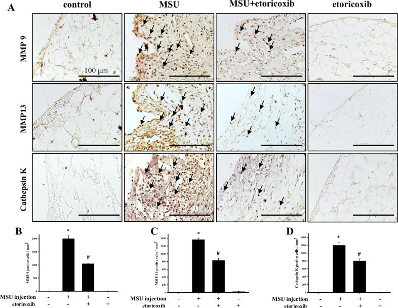
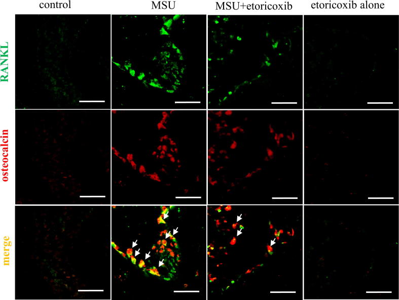
Deposition of monosodium urate (MSU) crystals in the joint or synovium is the major factor in Gouty arthritis (GA). The clinical features of chronic and recurrent GA include pain and the subsequent development of chronic tophaceous GA with multiple tophi deposits accompanied by osteolysis. The majority of previous animal studies have focused on MSU-induced acute GA without making observations regarding osteolysis. In the study, intra-articular injections of MSU into the knee (2 times/week for 10 weeks) was used to induce chronic and recurrent attacks of GA that in turn induced progressive osteolysis. Moreover, we also evaluated whether the clinical, nonsteroidal anti-inflammatory drug (NSAID) etoricoxib attenuated the osteoclastogenesis of progressive osteolysis. The knee morphometry and the expression of osteoclastogenesis-related proteins (cathepsin K and matrix metalloproteinase-9 and -13) in the knee were examined by micro-CT and immunohistochemistry, respectively. Results showed that oral etoricoxib not only significantly attenuated the nociceptive behaviors of the rats but that it also inhibited the expression of osteoclastogenesis-related proteins in their knee joints in chronic and recurrent attacks of GA. Our findings thus suggest that NSAIDs not only inhibit nociception but also prevent the progression of osteolysis in chronic and repeated attacks of GA.

Keywords: Chronic, recurrent gouty arthritis; Micro-CT; Osteoclastogenesis; Rat model.


Conflict of interest statement

The authors declare that they have no known competing financial interests or personal relationships that could have appeared to influence the work reported in this paper.

Figures

None
Graphical abstract
Fig. 1
Fig. 1
MSU to induce chronic and recurrent attacks of GA model. Time courses of the effects of etoricoxib on MSU-induced knee swelling (A), thermal hyperalgesia (C), mechanical allodynia (D), and hind paw weight-bearing deficits (E) in chronic GA. Oral administration of etoricoxib reduced MSU-induced knee swelling in the GA model. Each value show as mean ± SEM for each group. Results demonstrate that MSU significantly induced nociceptive behaviors and knee swelling and that etoricoxib could significantly reduce those effects. (B) Representative macroscopic images of the knee from the control, MSU, MSU + etoricoxib, and etoricoxib alone groups. Scale bar = 1 cm. * p < 0.05 symbolize significant variation between MSU and control groups at each time point. # p < 0.05 symbolize significant variation between MSU + etoricoxib and MSU group at each time point.
Fig. 2
Fig. 2
Micro-CT scan on knee of MUS-induced chronic, recurrent attacks of GA model. (A) Representative three-dimensional renderings of surface of knee, sagittal views, and trabecular bone scanned by micro-computed tomography. Red arrow indicates apparent bone erosion on surface of knee. Quantitative analysis of BMD (B), subchondral plate thickness (C), trabecular number (D), trabecular separation (E), and trabecular thickness (F), and analysis ROI (red frame) from surface of knee and sagittal views, respectively. * p < 0.05 symbolize significant variation between MSU and control groups. # p < 0.05 symbolize significant variation between MSU + etoricoxib and MSU group. (For interpretation of the references to color in this figure legend, the reader is referred to the web version of this article.)
Fig. 3
Fig. 3
Histopathological evaluation of MUS-induced chronic, recurrent attacks of GA model. Representative hematoxylin/eosin staining images for synovial tissue, Safranin O/Fast green staining images for cartilage and bone, and TRAP staining images for osteoclast of the knee joint sections of the control, MSU, MSU + etoricoxib, and etoricoxib alone groups. Oral administration of etoricoxib reduced that MSU-induced inflammation, cartilage damage, and osteoclast formation in the chronic GA model. * indicates apparent cartilage and bone erosion. Arrows indicate TRAP-positive cell. (For interpretation of the references to color in this figure legend, the reader is referred to the web version of this article.)
Fig. 4
Fig. 4
Immunohistochemical staining of MMP-9, MMP-13, and cathepsin K in synovial tissue. (A) Positive cells are indicated in brown (arrows) in joint sections from the control, MSU, MSU + etoricoxib, and etoricoxib alone groups. Quantitative analysis results of MMP-9- (B), MMP-13- (C), and cathepsin K- (D) positive cells of the synovial tissue are shown. Each value show as mean ± SEM for each group. Scale bar = 100 μm. * p < 0.05 symbolize significant variation between MSU and the control groups. # p < 0.05 symbolize significant variation between MSU + etoricoxib and MSU group.
Fig. 5
Fig. 5
Immunhistochemical stain of MMP9 MMP13 and cathepsin K on cartilage of knee. (A) Positive cells is shown in red-brown (arrow) in ankle joint sections from control, MSU, MSU + etoricoxib groups and etoricoxib alone groups. (B) Quantitative analysis of MMP 9 (B), MMP 13 (C) and cathepsin K (D) positive cell on cartilage are shown. Each value show as mean ± SEM for each group. Scale bar = 100 μm. * p < 0.05 symbolize significant variation between MSU and the control groups. # p < 0.05 symbolize significant variation between MSU + etoricoxib and the MSU group. (For interpretation of the references to color in this figure legend, the reader is referred to the web version of this article.)
Fig. 6
Fig. 6
Immunofluorescence staining of RANKL and osteocalcin in bone marrow of subchondral bone marrow. Representative confocal microscopy images showing the localization of RANKL (green) and osteocalcin (red) in subchondral bone marrow of the control, MSU, MSU + etoricoxib, and etoricoxib alone groups. Colocalization is symbolize by yellow color and arrows. The results showed that RANKL is primarily colocalized with osteoblasts. Scale bars = 50 µm. (For interpretation of the references to color in this figure legend, the reader is referred to the web version of this article.)

Similar articles

Cited by

References

    1. Underwood M. Gout BMJ Clin Evid. 2015;2015:1120. - PMC - PubMed
    1. Ames B.N., Cathcart R., Schwiers E., Hochstein P. Uric acid provides an antioxidant defense in humans against oxidant- and radical-caused aging and cancer: a hypothesis. Proc Natl Acad Sci U S A. 1981;78:6858–6862. - PMC - PubMed
    1. Ragab G., Elshahaly M., Bardin T. Gout: An old disease in new perspective - a review. J Adv Res. 2017;8:495–511. - PMC - PubMed
    1. Tausche A.K., Richter K., Grassler A., Hansel S., Roch B., Schroder H.E. Severe gouty arthritis refractory to anti-inflammatory drugs: treatment with anti-tumour necrosis factor alpha as a new therapeutic option. Ann Rheum Dis. 2004;63:1351–1352. - PMC - PubMed
    1. Sundy J.S., Baraf H.S., Yood R.A., Edwards N.L., Gutierrez-Urena S.R., Treadwell E.L. Efficacy and tolerability of pegloticase for the treatment of chronic gout in patients refractory to conventional treatment: two randomized controlled trials. JAMA. 2011;306:711–720. - PubMed

LinkOut - more resources