. 2020 Apr 2:8:16.
doi: 10.1038/s41413-020-0091-6. eCollection 2020.

Dystrophic calcification and heterotopic ossification in fibrocartilaginous tissues of the spine in diffuse idiopathic skeletal hyperostosis (DISH)

Affiliations

Dystrophic calcification and heterotopic ossification in fibrocartilaginous tissues of the spine in diffuse idiopathic skeletal hyperostosis (DISH)

Dale E Fournier et al. Bone Res. .

Abstract

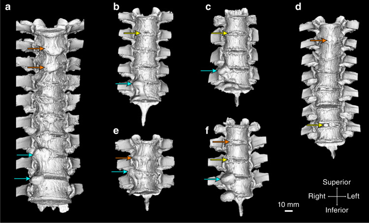
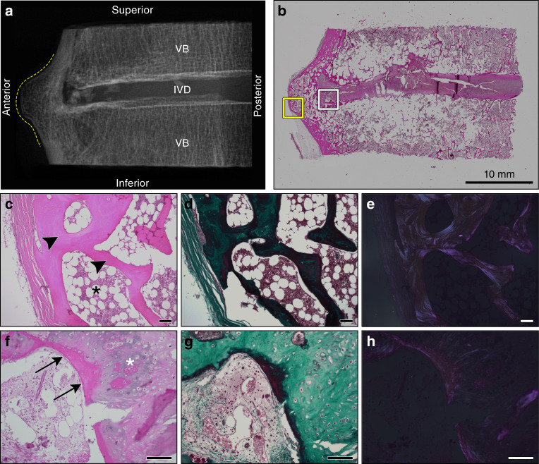
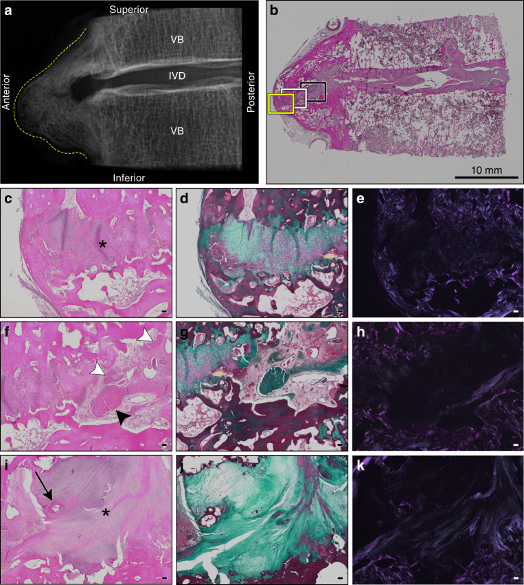
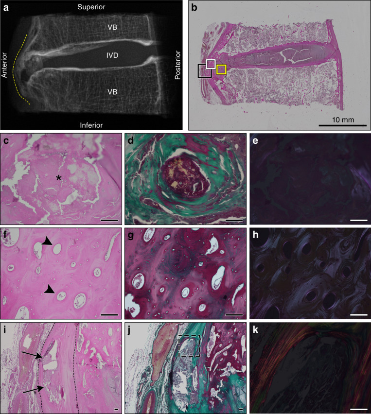
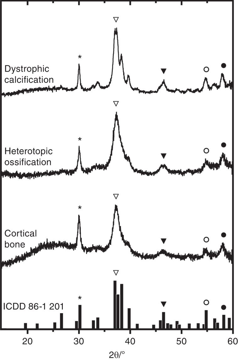
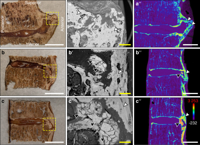
Diffuse idiopathic skeletal hyperostosis (DISH) is a prevalent noninflammatory spondyloarthropathy characterized by ectopic mineral formation along the anterolateral aspect of the vertebral column, yet little is known about its underlying pathogenesis. Our objective was to evaluate the histopathological features and composition of ectopic mineral within spinal tissues affected by DISH in humans. Thoracic spine segments from six embalmed cadaveric donors (one female and five males; median age 82 years) meeting the radiographic diagnostic criteria for DISH were evaluated using radiological, histological, and physical analyses. Overall, the histological features of ectopic mineralization at individual motion segments were heterogeneous, including regions of heterotopic ossification and dystrophic calcification. Heterotopic ossifications were characterized by woven and lamellar bone, multifocal areas of metaplastic cartilage, and bony bridges along the anterior aspect of the intervertebral disc space. Dystrophic calcifications were characterized by an amorphous appearance, a high content of calcium and phosphorus, an X-ray diffraction pattern matching that of hydroxyapatite, and radiodensities exceeding that of cortical bone. Dystrophic calcifications were found within the anterior longitudinal ligament and annulus fibrosus in motion segments both meeting and not meeting the radiographic criteria for DISH. In summary, our findings indicate that in DISH, ectopic mineral forms along the anterior aspect of the spine by both heterotopic ossification and dystrophic calcification of fibrocartilaginous tissues. Although both types of ectopic mineralization are captured by current radiographic criteria for DISH, dystrophic calcification may reflect a distinct disease process or an early stage in the pathogenesis of DISH.

Keywords: Bone; Pathogenesis.


Conflict of interest statement

Competing interestsThe authors declare no competing interests.

Figures

Fig. 1
Fig. 1
Three-dimensional isosurface renderings (anterior view) derived from microcomputed tomography (µCT) showing mineralized tissues (>310 Hounsfield units) of the spines included in the study. a 85-year-old male, thoracic vertebrae 4–11. b 77-year-old male, thoracic vertebrae 3–7. c 88-year-old male, thoracic vertebrae 7–9. d 72-year-old male, thoracic vertebrae 3–9. e 78-year-old male, thoracic vertebrae 4–6. f 86-year-old female, thoracic vertebrae 4–7. Arrows indicate the level of the intervertebral disc of the specific motion segments included in the study; yellow arrows—discontinous-patchy presentation, orange arrows—vertical presentation, and cyan arrows—horizontal presentation. The scale bar represents 10 mm
Fig. 2
Fig. 2
Histological appearance of a representative motion segment with horizontal presentation of ectopic mineralization associated with DISH. Images correspond to the specimen shown in Fig. 1b, T6-7. a Digital radiograph of the intact tissue prior to decalcification showing the localization of ectopic mineral within the motion segment (contoured by dotted yellow line). VB vertebral bone, IVD intervertebral disc. b Representative section stained with hematoxylin and eosin demonstrating the appearance of the intact section; scale bar represents 10 mm. The yellow box corresponds to an anterior region with features of heterotopic ossification imaged with a 10× objective (ce), while the white box corresponds to a transition region from heterotopic ossification to the underlying fibrocartilage annulus fibrosus imaged with a 20× objective (fh). c, f stained with hematoxylin and eosin; d, g stained with Masson’s trichrome; e, h the hematoxylin and eosin stained sections shown in c and f visualized with polarized light. Scale bars for ch represent 100 µm. ce Highlight features of heterotopic ossification along the anterior aspect of the motion segment, including compartmentalized bone marrow cavity (black asterisk) and organized lamellar bone (black arrowheads). fh Characteristics of primary woven bone (black arrows) and organized nests of chondrocytes (white asterisk) associated with endochondral ossification. All images are oriented as shown in a
Fig. 3
Fig. 3
Histological appearance of a representative motion segment with horizontal and discontinuous-patchy presentations of ectopic mineralization associated with DISH. Images correspond to the specimen shown in Fig. 1c, T8-9. a Digital radiograph of the intact tissue prior to decalcification showing the localization of ectopic mineral within the motion segment (contoured by dotted yellow line). VB vertebral bone, IVD intervertebral disc. b Representative section stained with hematoxylin and eosin demonstrating the appearance of the intact section; scale bar represents 10 mm. Yellow (ce), white (fh), and black boxes (ik) correspond to regions of fibrocartilage extending from the IVD positioned between mineralized outgrowths imaged with a 4× objective. c, f, i stained with hematoxylin and eosin; d, g, j stained with Masson’s trichrome; and e, h, and k the hematoxylin and eosin stained sections visualized with polarized light. Scale bars for ck represent 100 µm. ce Highlight the anterior-most portion of the fibrocartilaginous extension (asterisk). fh Reveal a unique transition zone marked by the presence of amorphous granular material (black arrowhead) and multifocal areas of fibrosis (white arrowheads). ik Display the fibrocartilage region (asterisk) adjacent to the native annulus fibrosus and a localized region of ossification (black arrow). All images are oriented as shown in a
Fig. 4
Fig. 4
Histological appearance of a representative motion segment with vertical presentation of ectopic mineralization associated with DISH. Images correspond to the specimen shown in Fig. 1a, T4–5. a Digital radiograph of the intact tissue prior to decalcification showing the localization of ectopic mineral within the motion segment (contoured by dotted yellow line). VB vertebral bone, IVD intervertebral disc. b Representative section stained with hematoxylin and eosin demonstrating the appearance of the intact section; scale bar represents 10 mm. The yellow box corresponds to the region within the fibrocartilage of the IVD with a discrete amorphous calcification imaged with a 20× objective (ce). The white box corresponds to a region of heterotopic ossification along the anterior aspect of the IVD imaged with a 20× objective (fh). The black box corresponds to the discrete calcification within the anterior longitudinal ligament imaged with 4× and 20× objectives (ik). c, f, i Stained with hematoxylin and eosin; d, g, j stained with Masson’s trichrome; and e, h, k the hematoxylin and eosin stained sections visualized with polarized light. Scale bars for ck represent 100 µm. ce Highlight an amorphous region of dystrophic calcification localized within the annulus fibrosus (asterisk) and characterized by a granular appearance and variable eosinophilic staining. fh A region of heterotopic ossification marked by organized osteons (black arrowheads). ik Reveal an area of dystrophic calcification (black arrows) localized within the anterior longitudinal ligament (contoured by black dotted lines). k Corresponds to the dotted outline in j. All images are oriented as shown in a
Fig. 5
Fig. 5
X-ray diffraction patterns from regions of ectopic mineralization associated with DISH. Shown are representative X-ray diffraction patterns from individual regions within sites of dystrophic calcification (n = 2 spines; 2 regions per deposit), heterotopic ossification (n = 3 spines; 2–4 regions per deposit), and unaffected cortical bone (3 spines; 1 region per segment) displayed in a stack plot. The results were matched to calcium-deficient hydroxyapatite [Ca8.8(PO4)6(OH)1.92] from the International Centre for Diffraction Data (powder diffraction file: 86–1 201), as indicated by the bar plot at the bottom and symbols
Fig. 6
Fig. 6
Characterization of the gross morphology and radiodensities of ectopic mineralizations associated with DISH. aaʹʹ Thoracic segment T9-10 from an 85-year-old male with heterotopic ossification (Fig. 1a), bbʹʹ thoracic segment T3-4 from a 72-year-old male with dystrophic calcification (Fig. 1f), and ccʹʹ thoracic segment T3-4 from an 85-year-old male with dystrophic calcification (Fig. 1a). ac The gross appearance of desiccated tissues with yellow dotted lines showing regions analyzed by scanning electron microscopy; white scale bars represent 10 mm. Corresponding scanning electron microscopy images; yellow scale bars represent 1 mm. aʹʹcʹʹ Pseudocolored radiodensity maps from matched µCT slices. Colors represent various radiodensities: purple (−232 to 310 Hounsfield units) corresponds to soft tissues; blue–green (310–2 750 Hounsfield units) corresponds to normal bone; and orange–red (2 750–3 253 Hounsfield units) corresponds to mineralizations exceeding the density of normal cortical bone. Topographical analysis of tissue sections by scanning electron microscopy highlighted the uniform, amorphous appearance of dystrophic calcifications (black arrowheads) within the intervertebral disc, which are distinct in appearance from adjacent ossifications (white arrowhead); white scale bars represent 10 mm. All images are oriented as shown in a

References

    1. Forestier J, Rotes-Querol J. Senile ankylosing hyperostosis of the spine. Ann. Rheum. Dis. 1950;9:321–330. doi: 10.1136/ard.9.4.321. - DOI - PMC - PubMed
    1. Resnick D, Niwayama G. Radiographic and pathologic features of spinal involvement in diffuse idiopathic skeletal hyperostosis (DISH) Radiology. 1976;119:559–568. doi: 10.1148/119.3.559. - DOI - PubMed
    1. Resnick D, Shaul SR, Robins JM. Diffuse idiopathic skeletal hyperostosis (DISH): forestier’s disease with extraspinal manifestations. Radiology. 1975;115:513–524. doi: 10.1148/15.3.513. - DOI - PubMed
    1. Mader R, Verlaan JJ, Buskila D. Diffuse idiopathic skeletal hyperostosis: clinical features and pathogenic mechanisms. Nat. Rev. Rheumatol. 2013;5:741–750. doi: 10.1038/nrrheum.2013.165. - DOI - PubMed
    1. Mata S, et al. A controlled study of diffuse idiopathic skeletal hyperostosis: clinical features and functional status. Med. (Baltim.) 1997;76:104–117. doi: 10.1097/00005792-199703000-00003. - DOI - PubMed