. 2020 Mar;3(1):22-33.
doi: 10.1093/pcmedi/pbz029. Epub 2019 Dec 30.

Potassium channels as potential drug targets for limb wound repair and regeneration

Affiliations

Potassium channels as potential drug targets for limb wound repair and regeneration

Wengeng Zhang et al. Precis Clin Med. 2020 Mar.

Abstract

Background: Ion channels are a large family of transmembrane proteins, accessible by soluble membrane-impermeable molecules, and thus are targets for development of therapeutic drugs. Ion channels are the second most common target for existing drugs, after G protein-coupled receptors, and are expected to make a big impact on precision medicine in many different diseases including wound repair and regeneration. Research has shown that endogenous bioelectric signaling mediated by ion channels is critical in non-mammalian limb regeneration. However, the role of ion channels in regeneration of limbs in mammalian systems is not yet defined.

Methods: To explore the role of potassium channels in limb wound repair and regeneration, the hindlimbs of mouse embryos were amputated at E12.5 when the wound is expected to regenerate and E15.5 when the wound is not expected to regenerate, and gene expression of potassium channels was studied.

Results: Most of the potassium channels were downregulated, except for the potassium channel kcnj8 (Kir6.1) which was upregulated in E12.5 embryos after amputation.

Conclusion: This study provides a new mouse limb regeneration model and demonstrates that potassium channels are potential drug targets for limb wound healing and regeneration.

Keywords: drug targets; gene expression; limb regeneration; potassium channels; wound healing.


Figures

Figure 1
Figure 1
Regeneration and wound healing of mouse embryonic limbs. (A and A’) A side view and a front view of a freshly wounded E12.5 embryonic hindlimb. The right hindlimb bud was amputated at the presumptive ankle level leaving an ovoid excisional surface without visible contraction of the skin tissues. (B and B’) A side view and a front view of a representative E12.5 embryo limb 24 hours after limb amputation. The wound is completely closed and two blastema-like structures form proximodistally at the anterior and posterior sides above the wound surface. (C) Immediately after the hindlimb was severed at the ankle level at an E15.5 embryo. The skin tissues around the wound are slightly contracted (red arrows). (D) 24 hours after limb amputation in an E15.5 embryo, the skin rounds up but the wound cannot close (red arrows). Note the dotted yellow line indicates the collection of tissues close to the wound surface.
Figure 2
Figure 2
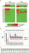
The relative expression level (REL) of 14 potassium channels during regeneration and wound healing. (A) Heatmap of the REL of potassium channels. Expression values for each gene at a row are colored from green (low) to red (high). As the expression levels of kcnj1 and kcnj8 (top two rows) are much higher than others, they are separated from other potassium channels to display a better color map. The leftmost column displays the gene names and the rightmost column shows the maximum changes of REL between all the different time points. Each time point was repeated once. The left half shows the REL at E12.5, and the right half at E15.5 embryos. (B) The REL of potassium channels at 0 hour after limb amputation. The values on the Y axis represent the REL between E12.5 and E15.5 limbs at 0 hour post amputation. The inset figure shows a more detailed view of the genes with a lower REL. kcnj1, kcna6, and kcnj11 are expressed at a much higher level, whereas kcnj8, kcnc4, and kcnk1 are expressed at a lower level in E12.5 limbs than E15.5 limbs. (C) Upregulation or downregulation of potassium channels at 24 hours versus 0 hour after limb amputation. The negative values denote downregulation, whereas the positive values represent upregulation. This shows that kcnj1, kcna6, and kcnb1 are downregulated at E12.5 limbs, and kcnh2 is downregulated at both E12.5 and E15.5 limbs, whereas kcnj8 is upregulated at E12.5 limbs by three times or more. All other values are lower than three times, indicating no remarkable changes.
Figure 3
Figure 3
kcna6/Kv1.6 expression after limb amputation in mouse embryos. (A) Real-time PCR and (B) representative Western blotting show that the relative expression level (REL) of kcna6 is high in E12.5 limbs and downregulated, while its expression is low in E15.5 limbs with no remarkable changes post wounding. (C) In situ hybridization shows that kcna6 is highly expressed at the wound surface immediately after limb amputation at E12.5 embryos (C, a) and is rapidly reduced after wound induction (C, b-d). There is no obvious expression at E15.5 embryonic limbs (C, e–h).
Figure 4
Figure 4
kcnj8/Kir6.1 expression after limb amputation in mouse embryos. (A) The real-time PCR shows that the relative expression level (REL) of kcnj8 is upregulated in E12.5 limbs whereas it is downregulated in E15.5 limbs after wounding. (B) Representative Western blotting indicates that kcnj8 is robustly induced after limb amputation at E12.5 embryos, but there is no detectable expression in E15.5 limbs, which may be a result of different post-translational modifications. (C) In situ hybridization shows that there is no obvious kncj8 expression at 0 hour (C, a) at E12.5 limbs (note the blue background in the whole embryo), whereas this is visible at the wound surface at 12 and 24 hours (C, c and d) after wound induction. There was no obvious staining in E15.5 wounded limbs (C, e–h).
Figure 5
Figure 5
kcnh2/Kv11.1 expression after limb amputation in mouse embryos. (A) Real-time PCR shows that the relative expression level (REL) of kcnh2 gene is high immediately after limb amputation and gradually downregulated afterwards in both E12.5 and E15.5 mouse limbs. (B) Representative Western blotting demonstrates that kcnh2 protein is downregulated after wound induction in both E12.5 and E15.5 limbs. But the kcnh2 protein is much lower at E15.5 limbs than E12.5, although the mRNA level is a little higher at E15.5 as shown in Fig. 4A at 0 hour. This is possibly a result of different post-translational modification of kcnh2 protein in E12.5 and E15.5 limbs. (C) In situ hybridization shows that kcnh2 is expressed at the wound surface (C, a) and reduced with time post wounding in E12.5 limbs (C, b-d). In E15.5 limbs, it is highly expressed mostly in the skin tissues of the wound surface (C, e and e’, a side and wound surface view) and downregulated during wound healing (C, f–h).

Similar articles

Cited by

References

    1. Alexander SPH, Mathie A, Peters JA, et al. . The concise guide to pharmacology 2019/20: Ion channels. Br J Pharmacol 2019;176Suppl 1:S142–S228. doi: 10.1111/bph.14749. - DOI - PMC - PubMed
    1. Levin M, Selberg J, Rolandi M. Endogenous bioelectrics in development, cancer, and regeneration: drugs and bioelectronic devices as electroceuticals for regenerative medicine. iScience 2019;22:519–33. doi: 10.1016/j.isci.2019.11.023. - DOI - PMC - PubMed
    1. Abdul Kadir L, Stacey M, Barrett-Jolley R. Emerging roles of the membrane potential: action beyond the action potential. Front Physiol 2018;9:1661. doi: 10.3389/fphys.2018.01661 - DOI - PMC - PubMed
    1. Haustrate A, Hantute-Ghesquier A, Prevarskaya N, et al. . Monoclonal antibodies targeting ion channels and their therapeutic potential. Front Pharmacol 2019;10:606. doi: 10.3389/fphar.2019.00606. - DOI - PMC - PubMed
    1. Weise-Cross L, Resta TC, Jernigan NL. Redox regulation of ion channels and receptors in pulmonary hypertension. Antioxid Redox Signal 2019;31:898–915. doi: 10.1089/ars.2018.7699. - DOI - PMC - PubMed

LinkOut - more resources