. 2020 Mar;3(1):34-43.
doi: 10.1093/pcmedi/pbaa001. Epub 2020 Jan 20.

Double negative T cells, a potential biomarker for systemic lupus erythematosus

Affiliations

Double negative T cells, a potential biomarker for systemic lupus erythematosus

Jessy J Alexander et al. Precis Clin Med. 2020 Mar.

Abstract

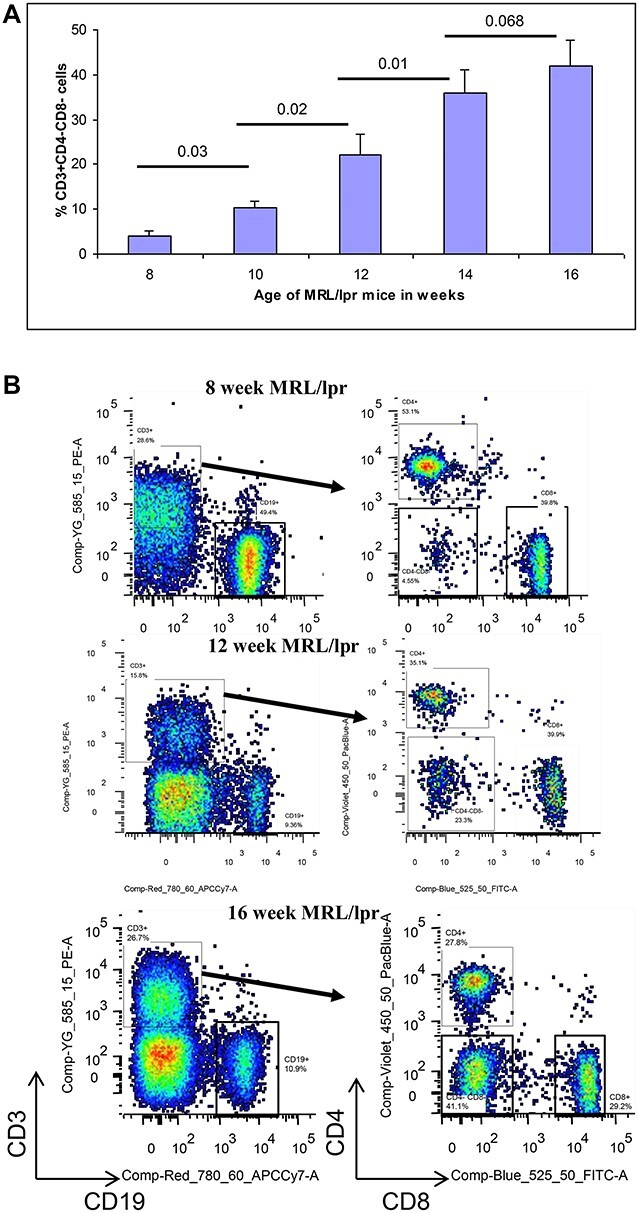
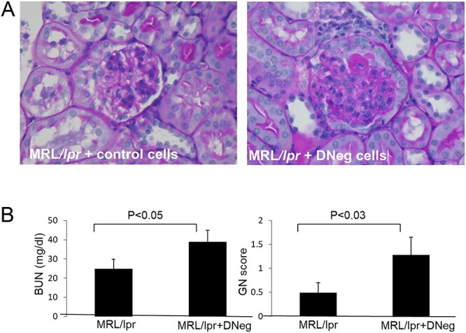
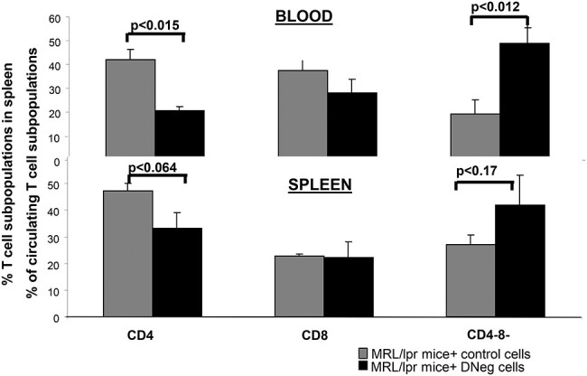
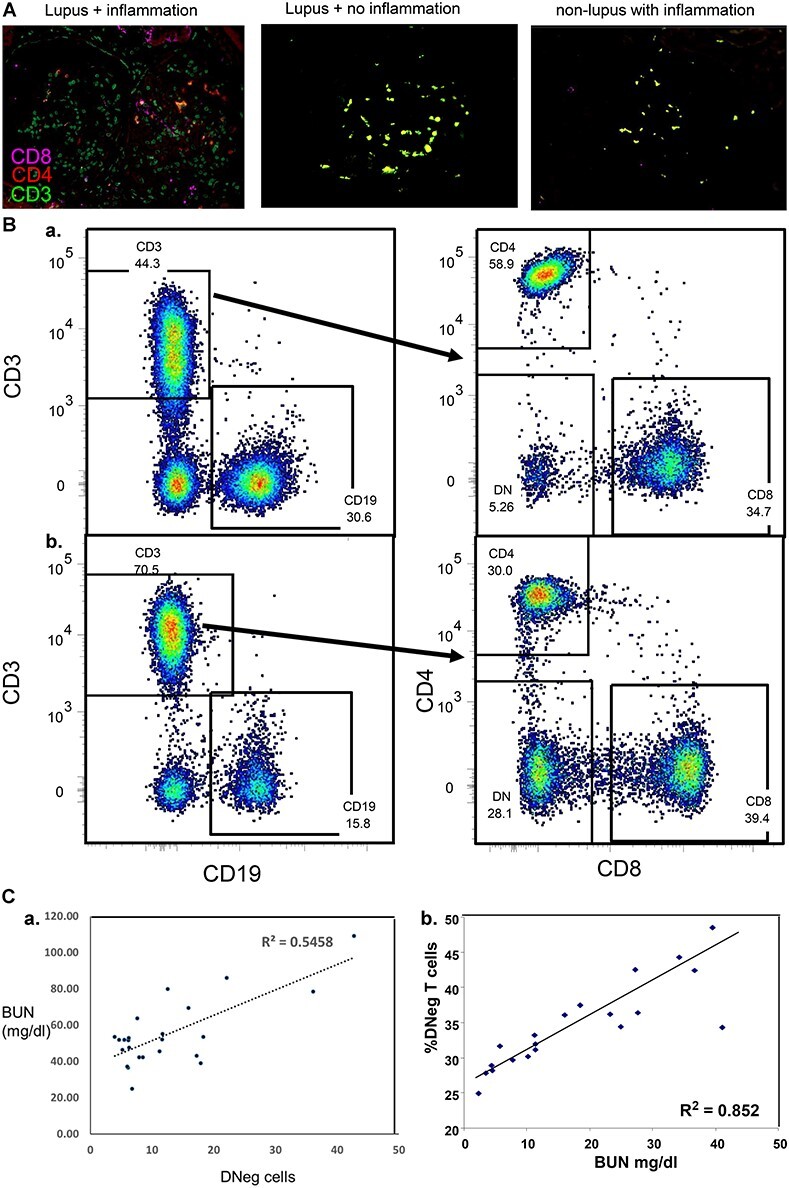
Systemic lupus erythematosus (SLE) is an autoimmune disease that is a challenge to diagnose and treat. There is an urgent need for biomarkers to help define organ involvement, and more effective therapies. A unique population of T cells, the CD3+CD4-CD8- (DNeg) cells, is significantly increased in lupus patients. Twenty-seven cases (53%) of pediatric SLE patients had elevated DNeg cells in their peripheral blood, which correlated with kidney function (R2 = 0.54). Significant infiltration of DNeg cells was observed in both adult and pediatric lupus kidneys by immunofluorescence. For the first time, this study provides direct evidence that DNeg cells facilitate kidney injury in preclinical 8-week-old MRL/lpr lupus mice. In lupus mice, the increase in DNeg cells tracked with worsening disease and correlated with kidney function (R2 = 0.85). Our results show that DNeg cells per se can cause kidney dysfunction, increase in number with increase in disease pathology, and could serve as a potential biomarker.

Keywords: CD3+CD4−CD8− T cells; glomerulonephritis; inflammation; lupus.


Figures

Figure 1
Figure 1
T cell population in blood from preclinical (8 weeks) to diseased (16 weeks) by FACS analysis. Flow cytometry was performed on peripheral blood mononuclear cells obtained from MRL/lpr mice from preclinical (8 weeks) to diseased (16 weeks) of age. At each time point, blood was obtained from six animals. Cells were stained with the following mAbs: Brilliant Violet 421 αCD3 (145C211), APC-Cy7 αCD19 (6D5), Alexa 647 αCD8 (KT15), FITC αCD4 (W3/25). They were analyzed for different T cell populations. (A) The T cell population was expressed as a percentage of gated CD3-positive cells. The percentage of CD3+CD4-CD8- cells increased with increasing age in MRL/lpr mice. (B) Representative data from time points 8, 12, and 16 weeks, n = 6 in each group.
Figure 2
Figure 2
Imagestream analysis of CD25 and FoxP3 on T cells from spleen and blood from MRL/lpr mice. Cells from spleen and blood were subjected to analysis by ImageStream 100 instrument (AMNIS Corporation, Seattle, WA). During ImageStream data acquisition, single cells are first separated from debris or multicellular events and composite images of single cell fluorescence intensities are given. Cells were stained with Brilliant Violet 421 αCD3 (145C211), APC-Cy7 αCD19 (6D5), Alexa Fluor 647 αCD8 (KT15), FITC αCD4 (W3/25), αCD25, and αFoxP3. CD25 was localized on the surface of CD4 cells and FoxP3 was cytosolic as expected. However, CD25 and FoxP3 were not expressed by either CD8 or CD4-CD8- cells.
Figure 3
Figure 3
Effect of adoptive transfer of DNeg cells on kidneys in young MRL/lpr mice. Representative periodic acid-Schiff–stained kidneys from 8-week-old MRL-lpr mice that received DNeg cells from 20-week MRL/lpr mice (experimental) or splenic cells from 8-week MRL/lpr mice (control) (A) and tissues harvested 5 weeks later. Increased pathology GN scores (B) with increased infiltration of inflammatory cells is seen in the glomeruli of the experimental mice compared to controls. Original magnifications, ×600. In line with the histology, kidney function assessed by BUN (B) was worsened in the experimental mice, P < 0.05.
Figure 4
Figure 4
Adoptive transfer of DNeg cells on CD4 and CD8 T cell populations in MRL/lpr mice. Single cell suspensions of spleen and blood depleted of RBCs were simultaneously stained for 1 hour with the following mAbs: Brilliant Violet 421 αCD3 (145C211), APC-Cy7 αCD19 (6D5), Alexa Fluor 647 αCD8 (KT15), FITC αCD4 (W3/25) and analyzed by flow cytometry. The T cell population was expressed as a percentage of gated CD3-positive cells of total splenocytes. Mice that received DNeg cells from aged MRL/lpr mice showed significant decrease in CD4+ and increase in DNeg cells, while CD8+ cells remained the same both in circulation and spleen compared to control mice (n = 3 in each group).
Figure 5
Figure 5
(A) DNeg cells are increased in kidneys from lupus patients. Representative confocal images of kidney biopsy specimens obtained from both adult and pediatric lupus were deparaffinized and stained with antibodies (CD3, green; CD4, red; and CD8, magenta) are shown. Scale bars: 50 μm. Original magnification: ×400. Images are representative of three biological replicates. (B) Circulating DNeg cells in pediatric lupus patients are increased compared to controls. Single cell suspensions from RBC depleted blood of pediatric patients were simultaneously stained for 1 hour with mAbs for CD3, CD19, CD8, CD4 and analyzed by flow cytometry. The T cell population is expressed as a percentage of gated CD3-positive cells. Normal subjects vs. lupus subjects = 6.5 ± 0.99 vs. 10 ± 6.15. 53% of lupus subjects had increased (0.8% of parent T cell population). FACS images of (a) control and (b) lupus subjects (B). (C) Circulating DNeg cells correlate with kidney function. BUN and DNeg cells were assessed in the same cases. Correlation between DNeg cells and BUN was significant in pediatric lupus patients (R2 = 0.5) and in lupus mice (R2 = 0.852).

Similar articles

Cited by

References

    1. Carroll MC. The lupus paradox. Nat Genet 1998;19:3–4. doi: 10.1038/ng0598-3. - DOI - PubMed
    1. Tarr T, Derfalvi B, Gyori N, et al. . Similarities and differences between pediatric and adult patients with systemic lupus erythematosus. Lupus 2015;24:796–803. doi: 10.1177/0961203314563817. - DOI - PubMed
    1. Fanouriakis A, Boumpas DT, Bertsias GK. Pathogenesis and treatment of CNS lupus. Curr Opin Rheumatol 2013;25:577–83. doi: 10.1097/BOR.0b013e328363eaf1. - DOI - PubMed
    1. Konya C, Paz Z, Tsokos GC. The role of T cells in systemic lupus erythematosus: an update. Curr Opin Rheumatol 2014;26:493–501. doi: 10.1097/BOR.0000000000000082. - DOI - PubMed
    1. Anand A, Dean GS, Quereshi K, et al. . Characterization of CD3+ CD4- CD8- (double negative) T cells in patients with systemic lupus erythematosus: activation markers. Lupus 2002;11:493–500. doi: 10.1191/0961203302lu235oa. - DOI - PubMed

LinkOut - more resources