. 2020 Mar 9;11(2):229-240.
doi: 10.14336/AD.2019.0510. eCollection 2020 Apr.

Ketogenic Diet Ameliorates Cardiac Dysfunction via Balancing Mitochondrial Dynamics and Inhibiting Apoptosis in Type 2 Diabetic Mice

Affiliations

Ketogenic Diet Ameliorates Cardiac Dysfunction via Balancing Mitochondrial Dynamics and Inhibiting Apoptosis in Type 2 Diabetic Mice

Yongzheng Guo et al. Aging Dis. .

Abstract

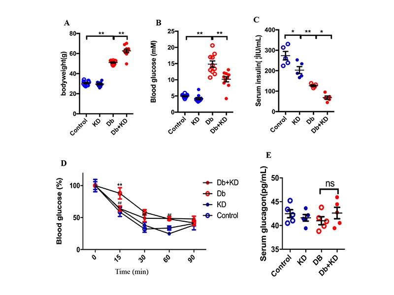
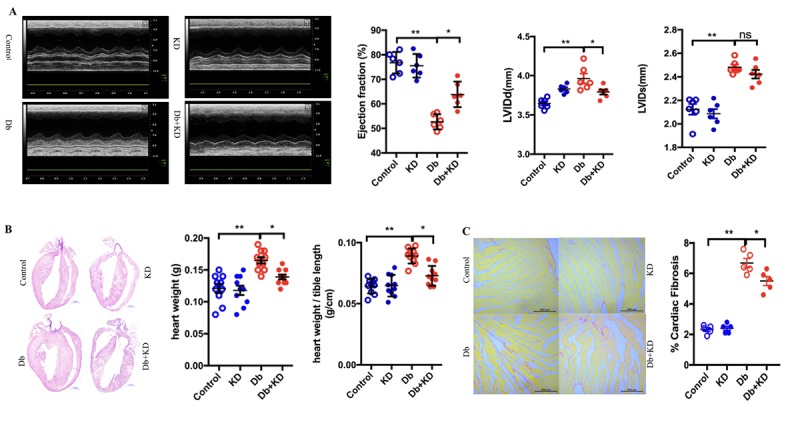
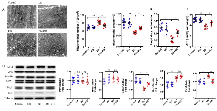
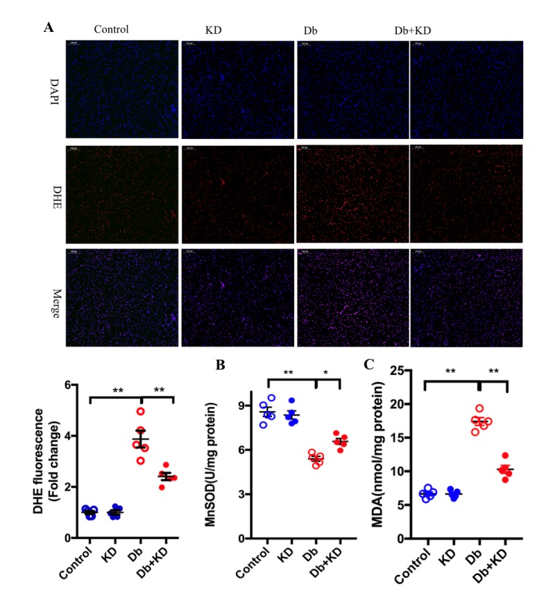
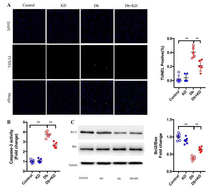
The ketogenic diet (KD) has been widely used in clinical studies and shown to hace an anti-diabetic effect, but the underlying mechanisms have not been fully elaborated. Our aim was to investigate the effects and the underling mechanisms of the KD on cardiac function in db/db mice. In the present study, db/db mice were subjected to a normal diet (ND) or KD. Fasting blood glucose, cardiac function and morphology, mitochondrial dynamics, oxidative stress, and apoptosis were measured 8 weeks post KD treatment. Compared with the ND, the KD improved glycemic control and protected against diabetic cardiomyopathy in db/db mice, and improved mitochondrial function, as well as reduced oxidative stress were observed in hearts. In addition, KD treatment exerted an anti-apoptotic effect in the heart of db/db mice. Further data showed that the PI3K/Akt pathway was involved in this protective effect. Our data demonstrated that KD treatment ameliorates cardiac dysfunction by inhibiting apoptosis via activating the PI3K-Akt pathway in type 2 diabetic mice, suggesting that the KD is a promising lifestyle intervention to protect against diabetic cardiomyopathy.

Keywords: db/db mice; diabetic cardiomyopathy; ketogenic diet; lifestyle intervention.


Figures

Figure 1.
Figure 1.
The ketogenic diet improved glycemic control and insulin sensitivity in db/db mice. Body weight (A), fasting blood glucose level (B), serum insulin level (C), insulin sensitivity test (D), and glucagon level (E) of mice after 8 weeks of feeding the KD; n=5-10. Values are the mean ± SEM. Data were analyzed using two-way ANOVA, followed by unpaired t-tests. #, P<0.05. ##, P< 0.01 versus Control. *, P<0.05. **, P<0.01 versus Db. ns indicates no significance.
Figure 2.
Figure 2.
The ketogenic diet improved cardiac function and alleviated cardiac remodeling in db/db mice. A) Feeding the KD improved the cardiac function in db/db mice. Typical echocardiogram images are shown in the left panel. LV ejection fraction, LV internal dimension diastole (LVIDd), and LV internal dimension systole (LVIDs) are shown in the right and lower panels; n=6 mice from each group. B) Cardiac phenotypes in the mice after 2 months of feeding the KD. Four-chamber view cardiac sections stained with hematoxylin and eosin staining. Scale bar, 1 mm. Heart weight and heart weight-to-tibia length ratio are shown in the right panel; n=10 mice from each group. C) Cardiac fibrosis in mice after 2 months of feeding the KD. Sirius red staining. Scale bar, 200μm. Cardiac fibrosis quantification as a percentage of the red stained area vs. total area stained by Sirius red is shown in the right panel; n=5 mice from each group. Values are the mean ± SEM. Data were analyzed using two-way ANOVA, followed by unpaired t-tests. *, P<0.05. **, P<0.01. ns indicates no significance.
Figure 3.
Figure 3.
The ketogenic diet prevented mitochondrial fission and improved mitochondrial function in the myocardium of db/db mice. A) Cardiac mitochondrial morphology in myocardial samples. Typical images obtained from electron microscopy are shown in the left panel. Scale bar, 1 μm. Average mitochondrial size and mitochondrial number are shown in the right panel; n=6 mice from each group. B) Mitochondrial respiratory control ration; n=5 mice from each group. C) ATP levels in the heart of the mice; n=5 mice from each group. D) The content of mitochondrial dynamics-related proteins in myocardial samples. Statistical results are shown in the right panel; n=6 mice from each group. Values are the mean ± SEM. Data are analyzed using two-way ANOVA, followed by unpaired t-tests. *, P<0.05. **, P<0.01. ns indicates no significance.
Figure 4.
Figure 4.
The ketogenic diet suppressed oxidative stress in the heart of db/db mice. A) Representative fluorescence microscopic image of DHE staining (red fluorescence) and DAPI-staining (blue fluorescence). Results derived from 5 different mice are shown below. Scale bar, 100 μm. Feeding the KD increased the activity of MnSOD (B) and decreased the content of MAD (C) in the myocardium of db/db mice; n=6 mice from each group. Values are the mean ± SEM. Data were analyzed using two-way ANOVA, followed by unpaired t-tests. *, P<0.05. **, P<0.01 versus Control.
Figure 5.
Figure 5.
The ketogenic diet attenuated cardiocyte apoptosis in db/db mice. A) Top representative TUNEL-stained (green fluorescence) and DAPI-stained (blue fluorescence) photomicrographs. Scale bar, 50 μm. Results derived from 5 different mice are shown in the right panel. B) Feeding the KD decreased caspase-3 activity. C) Feeding the KD increased the ratio of Bcl-2/Bax in the myocardium of db/db mice; n=6 mice from each group. Values are the mean ± SEM. Data were analyzed using two-way ANOVA, followed by unpaired t-tests. *, P<0.05. **, P<0.01.
Figure 6.
Figure 6.
BHB attenuated high glucose induced apoptosis in cardiomyocytes through the PI3K/Akt pathway. A) The KD increased the cardiac contents of PI3K and the ration of p-Akt/Akt in the myocardium of db/db mice. The typical images of western blot images are shown in the left panel. The statistical results are shown in the right panel; n=6 mice from each group. B) Effects of BHB on increasing the ration of Bcl2/Bax and p-Akt/Akt were blunted by the PI3K/Akt inhibitor wortmannin in cardiomyocytes exposed to high glucose; n=6. C) BHB decreased caspase-3 activity in cardiomyocytes exposed to high glucose; n=6. D) BHB protected cardiomyocytes against high glucose-induced apoptosis; n=6. Values are the mean ± SEM. Data were analyzed using two-way ANOVA, followed by unpaired t-tests. *, P<0.05. **, P<0.01.

Similar articles

Cited by

References

    1. Wallia A and Molitch ME (2014). Insulin therapy for type 2 diabetes mellitus. JAMA, 311: 2315-25. - PubMed
    1. Korkmaz-Icoz S, Al SS, Radovits T, Li S, Brune M, Hegedus P, et al. (2016). Oral treatment with a zinc complex of acetylsalicylic acid prevents diabetic cardiomyopathy in a rat model of type-2 diabetes: activation of the Akt pathway. Cardiovasc Diabetol, 15: 75. - PMC - PubMed
    1. Joubert M, Manrique A, Cariou B, Prieur X (2018). Diabetes-related cardiomyopathy: The sweet story of glucose overload from epidemiology to cellular pathways. Diabetes Metab. - PubMed
    1. Fein FS, Cho S, Malhotra A, Akella J, VanHoeven KH, Sonnenblick EH, et al. (1991). Beneficial effects of diltiazem on the natural history of hypertensive diabetic cardiomyopathy in rats. J Am Coll Cardiol, 18: 1406-17. - PubMed
    1. Fein FS (1990). Diabetic cardiomyopathy. Diabetes Care, 13: 1169-79. - PubMed

LinkOut - more resources