Impaired Frontoparietal Connectivity in Traumatic Individuals with Disorders of Consciousness: A Dynamic Brain Network Analysis
- PMID: 32257543
- PMCID: PMC7069467
- DOI: 10.14336/AD.2019.0606
Impaired Frontoparietal Connectivity in Traumatic Individuals with Disorders of Consciousness: A Dynamic Brain Network Analysis
Abstract
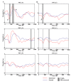
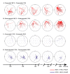
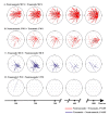
Recent advances in neuroimaging have demonstrated that patients with disorders of consciousness (DOC) may retain residual consciousness through activation of a complex functional brain network. However, an understanding of the hierarchy of residual consciousness and dynamic network connectivity in DOC patients is lacking. This study aimed to investigate residual consciousness and the dynamics of neural processing in DOC patients. We included 42 patients with DOC, categorized by aetiology. Event-related potentials combined with time-varying electroencephalography networks were used to probe affective consciousness in DOC and examine the related network mechanisms. The results showed an obvious frontal P3a component among patients in minimally conscious state (MCS), while a prominent N1 was observed in unresponsive wakefulness syndrome (UWS). No late positive potential (LPP) was detected in these patients. Next, we divided the results by aetiology. Patients with nontraumatic injury presented an obvious frontal P3a response compared to those with traumatic injury. With respect to the dynamic network mechanism, patients with UWS, both with and without trauma, exhibited impaired frontoparietal network connectivity during the middle to late emotion processing period (P3a and LPP). Surprisingly, unconscious post-traumatic patients had an evident deficit in top-down connectivity. This, it appears that early automatic sensory identification is preserved in UWS and that exogenous attention was preserved even in MCS. However, high-level cognitive abilities were severely attenuated in unconscious patients. We also speculate that reduced frontoparietal connectivity may be useful as a biomarker to distinguish patients in an MCS from those with UWS given the same aetiology.
Keywords: consciousness; event-related potentials; frontoparietal network; traumatic brain injury.
Copyright: © 2020 Wu et al.
Figures


References
-
- Machado C (2002). The minimally conscious state: definition and diagnostic criteria. Neurology, 59: 1473, 1473-1474. - PubMed
-
- Di HB, Yu SM, Weng XC, Laureys S, Yu D, Li JQ, et al. (2007). Cerebral response to patient's own name in the vegetative and minimally conscious states. Neurology, 68:895-899. - PubMed
-
- Fischer C, Luaute J, Morlet D (2010). Event-related potentials (MMN and novelty P3) in permanent vegetative or minimally conscious states. Clin Neurophysiol, 121: 1032-1042. - PubMed
LinkOut - more resources
Full Text Sources
