. 2020 Mar 4:10:8.
doi: 10.25259/JCIS_5_2020. eCollection 2020.

Computed Tomography-based Diagnosis of Post-laparoscopic Sleeve Gastrectomy Gastric Leak

Affiliations

Computed Tomography-based Diagnosis of Post-laparoscopic Sleeve Gastrectomy Gastric Leak

Sultan R Alharbi. J Clin Imaging Sci. .

Abstract

Objective: Gastric leak post-laparoscopic sleeve gastrectomy may appear as a variety of computed tomography (CT) findings. We aimed to review the various CT findings sensitivity and specificity in confirmed cases of gastric leak.

Materials and methods: A retrospective review was performed for all patients who underwent sleeve gastrectomy, CT, and endoscopy for suspected leak between 2011 and 2018. All patients with positive CT findings for gastric leak were included in the study.

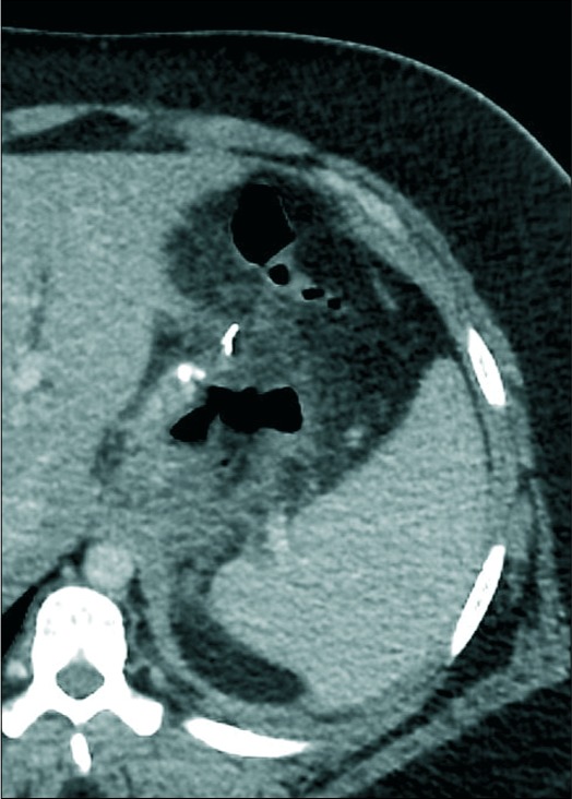
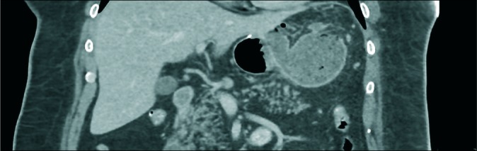
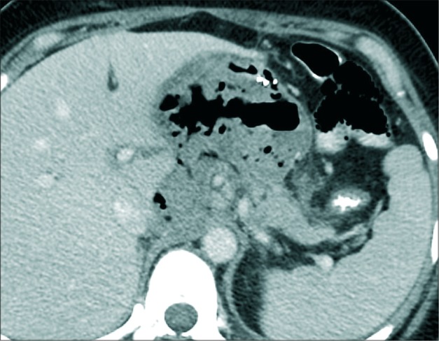
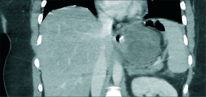
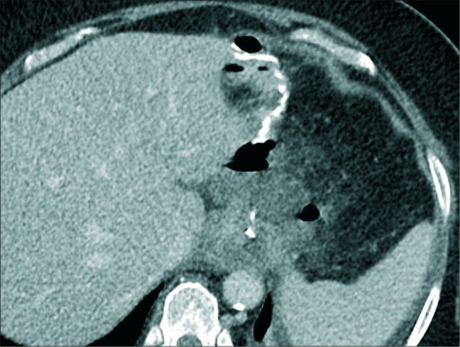
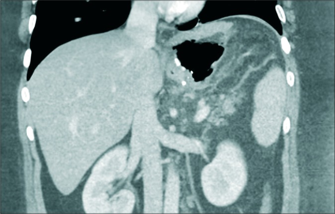
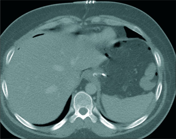
Results: A total of 152 consecutive patients underwent CT for suspected post-sleeve gastrectomy gastric leak. Out of 152 patients, 88 had positive CT findings for gastric leak and underwent endoscopy. The CT findings sensitivity and specificity of perigastric collection without oral contrast leak were 61% and 88.8%, oral contrast leak were 28% and 100%, and gas leak were 10% and 77.7%, respectively.

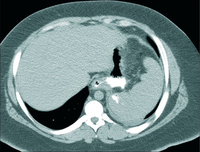
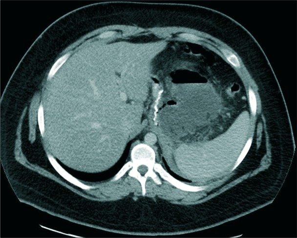
Conclusions: Perigastric fluid collection without contrast leak and with variable wall enhancement and gas content is the most common CT findings of post-sleeve gastrectomy gastric leak. Among various CT findings, it exhibits good diagnostic accuracy with 61% sensitivity and 88.8% specificity.

Keywords: Computed tomography; Gastric leak; Laparoscopic sleeve gastrectomy.


Conflict of interest statement

There are no conflicts of interest.

Figures

Figure 1:
Figure 1:
A 29-year-old female patient presented with abdominal pain, enhanced axial computed tomography image showing a normal post-operative gastric sleeve filled with oral contrast. No extraluminal gas or contrast. No perigastric fluid collection.
Figure 2:
Figure 2:
A 25-year-old male patient presented with repeated vomiting, enhanced axial computed tomography scan image showing oral contrast leak with gas contrast level.
Figure 3:
Figure 3:
A 34-year-old female patient presented with the left shoulder and chest pain, enhanced computed tomography scan image showing a gas leak only adjacent to the proximal gastric ruminant pouch.
Figure 4:
Figure 4:
A 19-year-old male patient presented with fever, enhanced axial computed tomography scan image showing perigastric collection without oral contrast leak with a gas fluid level and enhancing wall.
Figure 5:
Figure 5:
A 46-year-old male patient presented with abdominal pain and tachycardia, enhanced coronal computed tomography image showing perigastric fluid collection with a small amount of gas and well-defined enhancing wall.
Figure 6:
Figure 6:
A 26-year-old male patient presented with tachycardia, enhanced axial CT image showing large perigastric fluid collection with multiple air pockets displacing the gastric remnant anteriorly.
Figure 7:
Figure 7:
A 34-year-old male patient presented with tachycardia, enhanced axial computed tomography image showing perigastric fluid collection without an enhancing peripheral wall or gas.
Figure 8:
Figure 8:
A 60-year-old male patient presented with repeated vomiting, an enhanced axial computed tomography image showing perigastric fluid collection associated with fat stranding and a small pocket of air but no enhancing peripheral wall.
Figure 9:
Figure 9:
A 30-year-old male patient presented with the left shoulder pain and vomiting, enhanced coronal CT image showing extraluminal large gas pocket adjacent to gastric sleeve indicating a gas leak.
Figure 10:
Figure 10:
A 29-year-old male patient developed tachycardia and abdominal pain immediately after sleeve gastrectomy surgery, enhanced axial CT image showing post-operative expected multiple small air pockets in the non-dependent upper abdomen. No gas pocket has seen adjacent to gastric sleeve.

Similar articles

Cited by

References

    1. Tan JT, Kariyawasam S, Wijeratne T, Chandraratna HS. Diagnosis and management of gastric leaks after laparoscopic sleeve gastrectomy for morbid obesity. Obes Surg. 2010;20:403–9. doi: 10.1007/s11695-009-0020-7. - DOI - PubMed
    1. Abou Rached A, Basile M, El Masri H. Gastric leaks post sleeve gastrectomy: Review of its prevention and management. World J Gastroenterol. 2014;20:13904–10. doi: 10.3748/wjg.v20.i38.13904. - DOI - PMC - PubMed
    1. Buchwald H, Oien DM. Metabolic/bariatric surgery worldwide 2011. Obes Surg. 2013;23:427–36. doi: 10.1007/s11695-012-0864-0. - DOI - PubMed
    1. Himpens J, Dobbeleir J, Peeters G. Long-term results of laparoscopic sleeve gastrectomy for obesity. Ann Surg. 2010;252:319–24. doi: 10.1097/SLA.0b013e3181e90b31. - DOI - PubMed
    1. Gagner M, Deitel M, Erickson AL, Crosby RD. Survey on laparoscopic sleeve gastrectomy (LSG) at the fourth international consensus summit on sleeve gastrectomy. Obes Surg. 2013;23:2013–7. doi: 10.1007/s11695-013-1040-x. - DOI - PubMed

LinkOut - more resources