. 2020 Jan 8;9(2):66-71.
doi: 10.1007/s13691-019-00398-y. eCollection 2020 Apr.

Renal metastasis from intrahepatic cholangiocarcinoma

Affiliations

Renal metastasis from intrahepatic cholangiocarcinoma

Chihiro Matsumoto et al. Int Cancer Conf J. .

Abstract

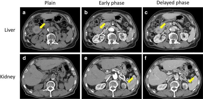
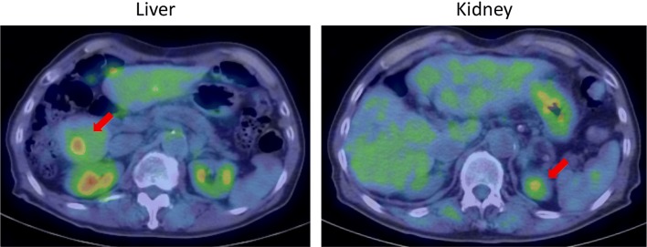
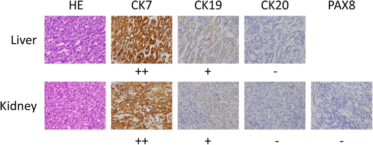
Metastases to the kidney are extremely rare and intrahepatic cholangiocarcinoma (ICC) is difficult to treat. In this study, we report a case of renal metastasis from ICC. A 72-year-old man who had been followed-up for chronic hepatitis C was diagnosed with ICC in the segment 8 and underwent S8 segmentectomy in 2014. During follow-up, the serum levels of carcinoembryonic antigen and carbohydrate antigen 19-9 were slightly elevated, and abdominal contrast-enhanced computed tomography revealed a low-density mass preceded by rim enhancement in the arterial phase measuring 1.5 × 1.5 cm in the segment 6, and a hypovascular mass measuring 2.2 × 2.0 cm in the upper pole of the left kidney in 2017. He underwent partial hepatectomy and partial nephrectomy. Based on postoperative histological findings combined with immunohistochemical analysis, the tumors both in the liver and kidney were diagnosed as recurrent ICC.

Keywords: Intrahepatic cholangiocarcinoma; Malignancy; Nephrectomy; Renal metastasis.


Conflict of interest statement

Conflict of interest The authors declare no conflicts of interest in association with this study.

Figures

Fig. 1
Fig. 1
Abdominal contrast-enhanced CT revealed a low-density mass preceded by rim enhancement in arterial phase measuring 1.5 × 1.5 cm in the segment 6 of the liver (ac), and hypovascular mass measuring 2.2 × 2.0 cm in the top of the left kidney (df)
Fig. 2
Fig. 2
PET-CT revealed that the both tumors in the liver and kidney showed significant uptake of fluorodeoxyglucose with standardized uptake rate max of 3.2 → 3.3 (liver) and 2.3 → 2.7 (kidney), respectively
Fig. 3
Fig. 3
On histological examination, the liver tumor revealed moderately differentiated tubular adenocarcinoma with confluent glandular and solid nest pattern. Immunohistochemical analysis showed that the tumor cells were diffusely positive for CK7, partially positive for CK19, whereas were negative for CK20, suggesting that the liver tumor was recurrent ICC. On the contrary, the histology of the renal tumor was solid, clear boundary and contained eosinophilic cytoplasm and revealed moderately differentiated tubular adenocarcinoma. Immunohistochemical analysis revealed that tumor cells were diffusely positive for CK7, partially positive for CK19, whereas were negative for CK20, PAX8 (Fig. 3). Based on these findings, the renal tumor was ultimately diagnosed as metastasis from ICC

References

    1. Ikai I, Arii S, Okazaki M, Okita K, Omata M, Kojiro M, Takayasu K, Nakanuma Y, Makuuchi M, Matsuyama Y, Monden M, Kudo M. Report of the 17th nationwide follow-up survey of primary liver cancer in Japan. Hepatol Res. 2007;37(9):676–691. doi: 10.1111/j.1872-034X.2007.00119.x. - DOI - PubMed
    1. Bridgewater J, Galle PR, Khan SA, Llovet JM, Park JW, Patel T, Pawlik TM, Gores GJ. Guidelines for the diagnosis and management of intrahepatic cholangiocarcinoma. J Hepatol. 2014;60(6):1268–1289. doi: 10.1016/j.jhep.2014.01.021. - DOI - PubMed
    1. Njei B. Changing pattern of epidemiology in intrahepatic cholangiocarcinoma. Hepatology. 2014;60(3):1107–1108. doi: 10.1002/hep.26958. - DOI - PubMed
    1. Hyder O, Hatzaras I, Sotiropoulos GC, Paul A, Alexandrescu S, Marques H, Pulitano C, Barroso E, Clary BM, Aldrighetti L, Ferrone CR, Zhu AX, Bauer TW, Walters DM, Groeschl R, Gamblin TC, Marsh JW, Nguyen KT, Turley R, Popescu I, Hubert C, Meyer S, Choti MA, Gigot JF, Mentha G, Pawlik TM. Recurrence after operative management of intrahepatic cholangiocarcinoma. Surgery. 2013;153(6):811–818. doi: 10.1016/j.surg.2012.12.005. - DOI - PMC - PubMed
    1. Abrams HL, Spiro R, Goldstein N. Metastases in carcinoma; analysis of 1000 autopsied cases. Cancer. 1950;3(1):74–85. doi: 10.1002/1097-0142(1950)3:1<74::AID-CNCR2820030111>3.0.CO;2-7. - DOI - PubMed

LinkOut - more resources