Methanol extract and fraction of Anchomanes difformis root tuber modulate liver mitochondrial membrane permeability transition pore opening in rats
- PMID: 32257891
- PMCID: PMC7103428
Methanol extract and fraction of Anchomanes difformis root tuber modulate liver mitochondrial membrane permeability transition pore opening in rats
Abstract
Objective: Extracts of Anchomanes difformis (AD) are used in folkloric medicine to treat several diseases and infections. However, their roles in mitochondrial permeability transition pore opening are not known.
Materials and methods: The viability of mitochondria isolated from Wistar rat liver used in this experiment, was assessed by monitoring their swelling amplitude in the absence of calcium and reversal of calcium-induced pore opening by spermine. The effects of methanol extract and fraction of A. difformis (MEAD and MFAD, respectively) on Mitochondrial Membrane Permeability Transition (MMPT) pore opening, ATPase activity, cytochrome c release and ferrous-induced lipid peroxidation were assessed spectrophotometrically. Phytochemical constituents of MEAD and MFAD were assessed using Gas Chromatography- Mass Spectrometry (GC-MS).
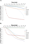
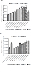
Results: The MEAD (10, 20, 40 and 80 μg/ ml) had no effect on MMPT pore opening in the absence of Ca2+, whereas MFAD at 80 μg/ml had a large amplitude pore opening effect. Both MEAD and MFAD reversed Ca2+-induced swelling with inhibition values of 18, 21, 24, 23% (for MEAD) and 41, 36, 35, and 26% (for MFAD) at 10, 20, 40 and 80 μg/ml, respectively. MFAD significantly enhanced F1F0 ATPase activity and caused cytochrome c release. Both MEAD and MFAD significantly inhibited ferrous-induced lipid peroxidation by 33.0, 64.0, 66, and 75% (for MEAD) and 24, 25, 30, and 45% (for MFAD), respectively. The GC-MS results revealed the presence of squalene as one of the major constituents of MEAD.
Conclusion: These findings suggest that MFAD can be used to induce cell death via mitochondrial permeability transition in isolated rat liver. Inhibition of lipid peroxidation by MEAD and MFAD showed that the pore opening effect of the extract and fraction was not mediated via peroxidation of mitochondrial membrane lipids.
Keywords: Anchomanes difformis; Cytochrome c; Lipid peroxidation; Mitochondrial permeability Transition pore opening; Phytoconstituents Mitochondrial ATPase.
Figures


Similar articles
-
Effects of extracts of the leaves of Brysocarpus coccineus on rat liver mitochondrial membrane permeability transition (MMPT) pore.Afr J Med Med Sci. 2012 Dec;41 Suppl:125-32. Afr J Med Med Sci. 2012. PMID: 23678647
-
Effects of methanolic and chloroform extracts of leaves of Alstonei boonei on rat liver mitochondrial membrane permeability transition pore.Afr J Med Med Sci. 2010 Dec;39 Suppl:109-16. Afr J Med Med Sci. 2010. PMID: 22416652
-
Modulation of opening of rat liver mitochondrial membrane permeability transition pore by different fractions of the leaves of Cnestis ferruginea. D.C.Afr J Med Med Sci. 2012 Dec;41 Suppl:157-69. Afr J Med Med Sci. 2012. PMID: 23678652
-
Induction of mitochondrial-dependent apoptosis, activation of mitochondrial ATPase and cytochrome c release by the methanol extract of Funtumia elastica stem bark.J Complement Integr Med. 2025 Apr 14. doi: 10.1515/jcim-2024-0326. Online ahead of print. J Complement Integr Med. 2025. PMID: 40215108
-
Induction of rat hepatic mitochondrial membrane permeability transition pore opening by leaf extract of Olax subscorpioidea.Pharmacognosy Res. 2015 Jun;7(Suppl 1):S63-8. doi: 10.4103/0974-8490.157998. Pharmacognosy Res. 2015. PMID: 26109790 Free PMC article.
Cited by
-
Studies on the mitochondrial, immunological and inflammatory effects of solvent fractions of Diospyros mespiliformis Hochst in Plasmodium berghei-infected mice.Sci Rep. 2021 Mar 25;11(1):6941. doi: 10.1038/s41598-021-85790-6. Sci Rep. 2021. PMID: 33767260 Free PMC article.
-
Leaf Extracts of Anchomanes difformis Ameliorated Kidney and Pancreatic Damage in Type 2 Diabetes.Plants (Basel). 2021 Feb 5;10(2):300. doi: 10.3390/plants10020300. Plants (Basel). 2021. PMID: 33562428 Free PMC article.
References
-
- Alabi TD, Brooks N, Oguntibeju O. The potency of medicinal plants in the treatment and management of Diabetes mellitus. Apple Academy Press. First Edition Chapter 9. Medicinal activities of Anchomanes difformis and its potential in the treatment of diabetes mellitus and other diseases conditions: A review; pp. 219–235.
-
- Ankur R, Arti M, Seema R, Amarjeet D, Ashok K. Mitochondrial permeability transition pore: another review. Int Res J Pharm. 2012;3:106–108.
-
- Appaix F, Minatchy MN, Riva-Lavieille C, Olivares J, Antonsson B, Saks VA. Rapid spectrophotometric method for quantitation of cytochrome c release from isolated mitochondria or permeabilized cells revisited. Biochim Biophys Acta. 2000;1457:175–181. - PubMed
LinkOut - more resources
Full Text Sources
Miscellaneous
