

Tracking acute phase protein response during acute and chronic Toxoplasma gondii infection
- PMID: 32257894
- PMCID: PMC7081684
- DOI: 10.1186/s42826-019-0007-z
Tracking acute phase protein response during acute and chronic Toxoplasma gondii infection
Abstract
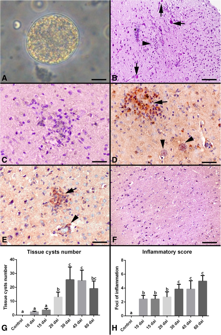
Toxoplasmosis is a disease caused by the protozoan Toxoplasma gondii, which occurs worldwide in mammals and birds. Brain is the primary target organ because Toxoplasma gondii is a ubiquitous intracellular parasite that causes most frequently life-threatening encephalitis in immunocompromised patients. Relation of tissue cysts number, histopathology score and acute phase proteins were investigated. In this study, 36 mice are infected with Me49 strain of Toxoplasma gondii. The control group has 6 healthy mice. After inoculation of Toxoplasma gondii, at 10., 15., 20., 30., 45., 60. days, 6 each mice euthanized after collection of blood samples. Hemopexin, haptoglobulin, macroglobulin, serum amyloid A and clusterin levels are determined by ELISA. Then, brain tissues were investigated histopathologically and lesions were scored. The average cyst numbers were determined by counting three samples (25 μl each) of each brain homogenate under light microscopy. Inflammatory reaction was observed on day 10 days after inoculation (d.a.i.) The lesions were characterized by perivascular mononuclear cell infiltration, focal mononuclear cell infiltration in the meninges, and glial proliferation. Tissue cysts were observed in all Toxoplasma gondii-infected groups. The highest lesion score was observed at 60 d.a.i. And the most tissue cyst number were on day 30. d.a.i. Serum levels of hemopexin, haptoglobulin, macroglobulin, serum amyloid A and clusterin were significantly higher than the control group on day 10-20., 10., 10-30., 10.,10-45 d.a.i., respectively. High level of acute phase proteins in mice on certain days infected with Toxoplasma gondii was exhibited a relationship between brain lesions and tissue cysts.
Keywords: Acute; Acute phase proteins; Chronic; Encephalitis; Toxoplasma gondii.
© The Author(s) 2019.
Conflict of interest statement
Competing interestsThe authors declare that they have no competing interests.
Figures

References
-
- Dubey JP, Beattie CP. Toxoplasmosis of animals and man. Boca Raton, Fla.: CRC Press; 1988. http://www.loc.gov/catdir/enhancements/fy0744/87026826-d.html.
-
- Atmaca HT, Kul O, Karakuş E, Terzi OS, Canpolat S, Anteplioğlu T. Astrocytes, microglia/macrophages, and neurons expressing toll-like receptor 11 contribute to innate immunity against encephalitic toxoplasma gondii infection. Neuroscience. 2014;269:184–191. doi: 10.1016/j.neuroscience.2014.03.049. - DOI - PubMed
-
- Dubey JP, Lindsay DS. Opportunistic infections: toxoplasma, Sarcocystis, and microsporidia. Boston, MA: Springer US; 2004. Biology of toxoplasma Gondii in cast and other animals; pp. 1–19.
LinkOut - more resources
Full Text Sources
