

Age-dependent changes of p53 and p63 immunoreactivities in the mouse hippocampus
- PMID: 32257908
- PMCID: PMC7081572
- DOI: 10.1186/s42826-019-0022-0
Age-dependent changes of p53 and p63 immunoreactivities in the mouse hippocampus
Abstract
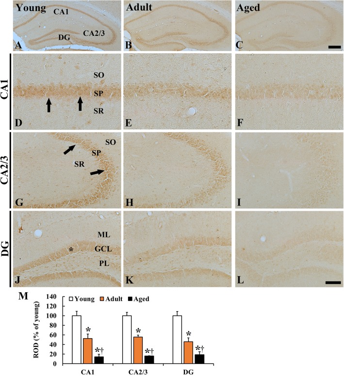
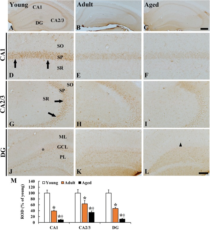
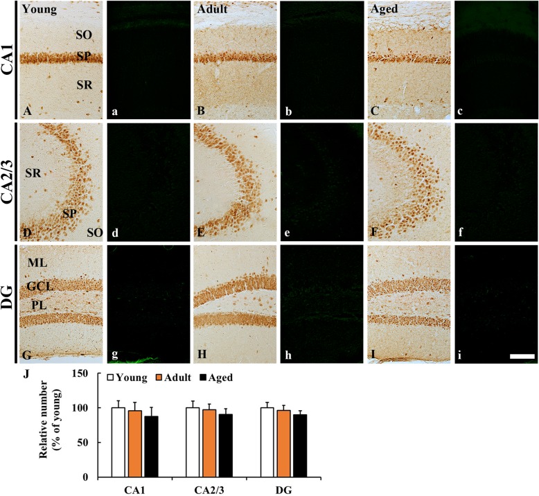
P53 and its family member p63 play important roles in cellular senescence and organismal aging. In this study, p53 and p63 immunoreactivity were examined in the hippocampus of young, adult and aged mice by using immunohistochemistry. In addition, neuronal distribution and degeneration was examined by NeuN immunohistochemistry and fluoro-Jade B fluorescence staining. Strong p53 immunoreactivity was mainly expressed in pyramidal and granule cells of the hippocampus in young mice. p53 immunoreactivity in the pyramidal and granule cells was significantly reduced in the adult mice. In the aged mice, p53 immunoreactivity in the pyramidal and granule cells was more significantly decreased. p63 immunoreactivity was strong in the pyramidal and granule cells in the young mice. p63 immunoreactivity in these cells was apparently and gradually decreased with age, showing that p63 immunoreactivity in the aged granule cells was hardly shown. However, numbers of pyramidal neurons and granule cells were not significantly decreased in the aged mice with normal aging. Taken together, this study indicates that there are no degenerative neurons in the hippocampus during normal aging, showing that p53 and p63 immunoreactivity in hippocampal neurons was progressively reduced during normal aging, which might be closely related to the normal aging processes.
Keywords: Aging process; Granule cells; Hippocampus; Mouse; Pyramidal neurons; p53; p63.
© The Author(s) 2019.
Conflict of interest statement
Competing interestsThe authors have no financial competing interest.
Figures
Similar articles
-
Delayed hippocampal neuronal death in young gerbil following transient global cerebral ischemia is related to higher and longer-term expression of p63 in the ischemic hippocampus.Neural Regen Res. 2015 Jun;10(6):944-50. doi: 10.4103/1673-5374.158359. Neural Regen Res. 2015. PMID: 26199612 Free PMC article.
-
p63 Expression in the Gerbil Hippocampus Following Transient Ischemia and Effect of Ischemic Preconditioning on p63 Expression in the Ischemic Hippocampus.Neurochem Res. 2015 May;40(5):1013-22. doi: 10.1007/s11064-015-1556-7. Epub 2015 Mar 18. Neurochem Res. 2015. PMID: 25777256
-
Ontogeny of calbindin immunoreactivity in the human hippocampal formation with a special emphasis on granule cells of the dentate gyrus.Int J Dev Neurosci. 2009 Apr;27(2):115-27. doi: 10.1016/j.ijdevneu.2008.12.004. Epub 2008 Dec 27. Int J Dev Neurosci. 2009. PMID: 19150647
-
Cyclin D1 immunoreactivity changes in CA1 pyramidal neurons and dentate granule cells in the gerbil hippocampus after transient forebrain ischemia.Neurol Res. 2011 Jan;33(1):93-100. doi: 10.1179/016164110X12714125204399. Epub 2010 Jun 11. Neurol Res. 2011. PMID: 20546683
-
Age-related biophysical alterations of hippocampal pyramidal neurons: implications for learning and memory.Ageing Res Rev. 2002 Apr;1(2):181-207. doi: 10.1016/s1568-1637(01)00009-5. Ageing Res Rev. 2002. PMID: 12039438 Review.
Cited by
-
Divergence of the PIERCE1 expression between mice and humans as a p53 target gene.PLoS One. 2020 Aug 3;15(8):e0236881. doi: 10.1371/journal.pone.0236881. eCollection 2020. PLoS One. 2020. PMID: 32745107 Free PMC article.
-
Comparison of age-dependent alterations in thioredoxin 2 and thioredoxin reductase 2 expressions in hippocampi between mice and rats.Lab Anim Res. 2021 Mar 6;37(1):11. doi: 10.1186/s42826-021-00088-y. Lab Anim Res. 2021. PMID: 33676586 Free PMC article.
References
LinkOut - more resources
Full Text Sources
Research Materials
Miscellaneous
