. 2019 Nov 21;36(2):167-173.
doi: 10.1007/s43188-019-00025-1. eCollection 2020 Apr.

Arylquin 1, a potent Par-4 secretagogue, induces lysosomal membrane permeabilization-mediated non-apoptotic cell death in cancer cells

Affiliations

Arylquin 1, a potent Par-4 secretagogue, induces lysosomal membrane permeabilization-mediated non-apoptotic cell death in cancer cells

Kyoung-Jin Min et al. Toxicol Res. .

Abstract

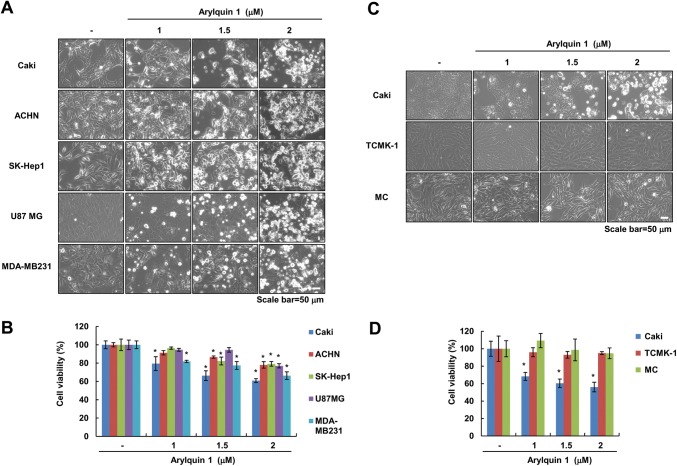
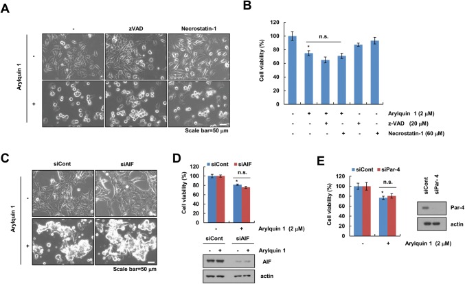
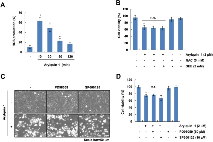
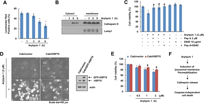
Arylquin 1, a small-molecule prostate-apoptosis-response-4 (Par-4) secretagogue, targets vimentin to induce Par-4 secretion. Secreted Par-4 binds to its receptor, 78-kDa glucose-regulated protein (GRP78), on the cancer cell surface and induces apoptosis. In the present study, we investigated the molecular mechanisms of arylquin 1 in cancer cell death. Arylquin 1 induces morphological changes (cell body shrinkage and cell detachment) and decreases cell viability in various cancer cells. Arylquin 1-induced cell death is not inhibited by apoptosis inhibitors (z-VAD-fmk, a pan-caspase inhibitor), necroptosis inhibitors (necrostatin-1), and paraptosis inhibitors. Furthermore, arylquin 1 significantly induces reactive oxygen species levels, but antioxidants [N-acetyl-l-cysteine and glutathione ethyl ester] do not inhibit arylquin 1-induced cell death. Furthermore, Par-4 knock-down by small interfering RNA confers no effect on cytotoxicity in arylquin 1-treated cells. Interestingly, arylquin 1 induces lysosomal membrane permeabilization (LMP), and cathepsin inhibitors and overexpression of 70-kDa heat shock protein (HSP70) markedly prevent arylquin 1-induced cell death. Therefore, our results suggest that arylquin 1 induces non-apoptotic cell death in cancer cells through the induction of LMP.

Keywords: Arylquin 1; Cell death; Lysosomal membrane permeabilization; Non-apoptotic cell death; Prostate‐apoptosis‐response‐4.


Conflict of interest statement

Conflict of interestThe authors declare no conflicts of interest.

Figures

Fig. 1
Fig. 1
Arylquin 1 induces cell death in various cancer cells. ad Cells were treated with the indicated concentrations of arylquin 1 for 24 h. Cell morphology was examined using interference light microscopy (a, c). Cell viability was determined using the XTT assay (b, d). The values in b, d represent the mean ± SEM from three independent samples. *p < 0.05 compared to the control. SEM standard error of the mean
Fig. 2
Fig. 2
Arylquin 1-induced cell death is independent of caspase, AIF, and Par-4. a, b Caki cells were pretreated with 20 μM z-VAD and 60 μM necrostatin-2 for 30 min, and then treated with 2 μM arylquin 1 for 24 h. c, d Caki cells were transiently transfected with control siRNA (siCont) and AIF siRNA (siAIF). After 24 h, Caki cells were treated with 2 μM arylquin 1 for 24 h. e Caki cells were transiently transfected with control siRNA (siCont) and Par-4 siRNA (siPar-4). After 24 h, Caki cells were treated with 2 μM arylquin 1 for 24 h. The cell morphology was examined using interference light microscopy (a, c). Cell viability was determined using the XTT assay (b, d, e). The protein expression levels of AIF, Par-4, and actin were determined by western blotting. The values in b, d, e represent the mean ± SEM from three independent samples. *p < 0.05 compared to the control
Fig. 3
Fig. 3
Reactive oxygen species and paraptosis are not involved in arylquin 1-induced cell death. a Caki cells were treated with 2 μM arylquin 1 for the indicated time periods. After treatment, cells were stained with H2DCFDA dye. Fluorescence was detected using flow cytometry. b Caki cells were pretreated with 5 mM NAC and 2 mM GEE for 30 min, and then cells were treated with 2 μM arylquin 1 for 24 h. Cell viability was determined using the XTT assay. c, d Caki cells were pretreated with 50 μM PD98059 and 10 μM SP600125 for 30 min, and then added with the 2 μM arylquin 1 for 24 h. Cell morphology was examined using interference light microscopy (c). Cell viability was determined using the XTT assay (d). The values in a, b, d represent the mean ± SEM from three independent samples. *p < 0.05 compared to the control
Fig. 4
Fig. 4
Arylquin 1 induces lysosomal membrane permeabilization-mediated cell death. a, b Caki cells were treated with 2 μM arylquin 1 for the indicated time periods, and then cells were incubated with the LysoTracker Red fluorescent dye. The fluorescence intensity was detected using flow cytometry (a). Cytosol and membrane fractions (lysosome-rich fraction) were prepared, and the protein expression levels of cathepsin D and Lamp1 were determined by western blotting (b). c Caki cells were pretreated with 2 μM pepstatin A (Pep A) and/or 10 μg/mL E64D for 30 min, and then added with 2 μM arylquin 1 for 24 h. Cell viability was determined using the XTT assay. d, e Caki/vector and Caki/HSP70 cells were treated with the indicated concentrations of arylquin 1 for 24 h. Cell morphology was examined using interference light microscopy (d). Cell viability was determined using the XTT assay (e). f Schematic diagram of arylquin 1-induced cell death in cancer cells. The protein expression levels of HSP70 and actin were determined by western blotting. The values in a, c, e represent the mean ± SEM from three independent samples. *p < 0.05 compared to the control. #p < 0.05 compared to the arylquin 1

Similar articles

Cited by

References

    1. Burikhanov R, Sviripa VM, Hebbar N, Zhang W, Layton WJ, Hamza A, Zhan CG, Watt DS, Liu C, Rangnekar VM. Arylquins target vimentin to trigger Par-4 secretion for tumor cell apoptosis. Nat Chem Biol. 2014;10:924–926. doi: 10.1038/nchembio.1631. - DOI - PMC - PubMed
    1. Sviripa VM, Burikhanov R, Obiero JM, Yuan Y, Nickell JR, Dwoskin LP, Zhan CG, Liu C, Tsodikov OV, Rangnekar VM, Watt DS. Par-4 secretion: stoichiometry of 3-arylquinoline binding to vimentin. Org Biomol Chem. 2016;14:74–84. doi: 10.1039/C5OB01980J. - DOI - PMC - PubMed
    1. Sells SF, Wood DP, Jr, Joshi-Barve SS, Muthukumar S, Jacob RJ, Crist SA, Humphreys S, Rangnekar VM. Commonality of the gene programs induced by effectors of apoptosis in androgen-dependent and -independent prostate cells. Cell Growth Differ. 1994;5:457–466. - PubMed
    1. Chakraborty M, Qiu SG, Vasudevan KM, Rangnekar VM. Par-4 drives trafficking and activation of Fas and Fasl to induce prostate cancer cell apoptosis and tumor regression. Can Res. 2001;61:7255–7263. - PubMed
    1. Burikhanov R, Zhao Y, Goswami A, Qiu S, Schwarze SR, Rangnekar VM. The tumor suppressor Par-4 activates an extrinsic pathway for apoptosis. Cell. 2009;138:377–388. doi: 10.1016/j.cell.2009.05.022. - DOI - PMC - PubMed