

Three-dimensional cone beam computed tomography analysis of temporomandibular joint response to the Twin-block functional appliance
- PMID: 32257934
- PMCID: PMC7093662
- DOI: 10.4041/kjod.2020.50.2.86
Three-dimensional cone beam computed tomography analysis of temporomandibular joint response to the Twin-block functional appliance
Abstract
Objective: To propose a three-dimensional (3D) method for evaluating temporomandibular joint (TMJ) changes during Twin-block treatment.
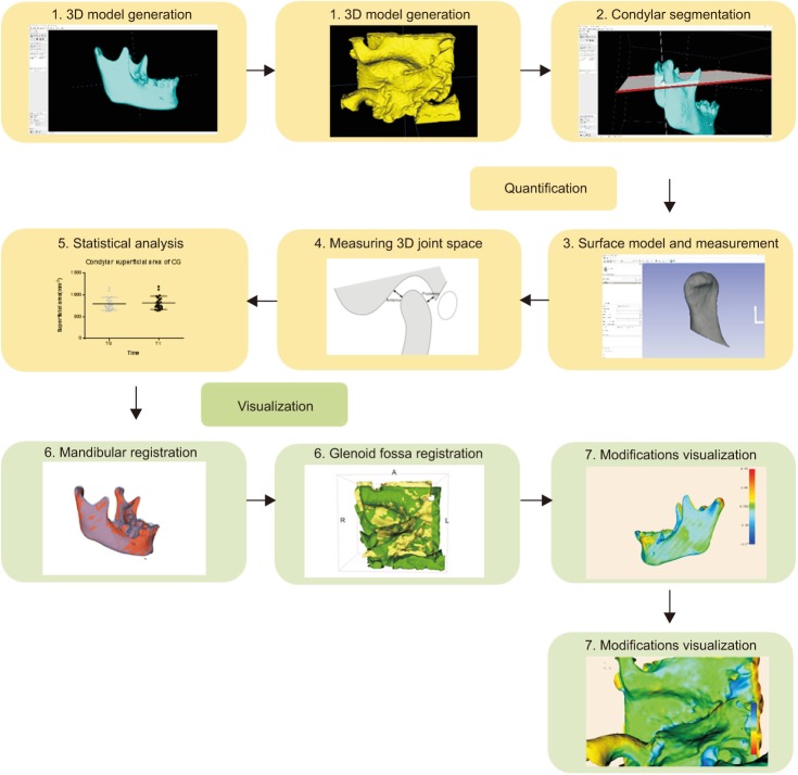
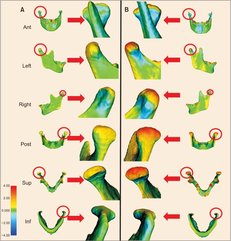
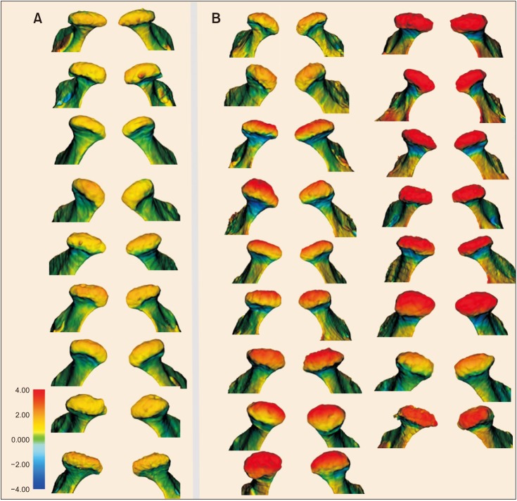
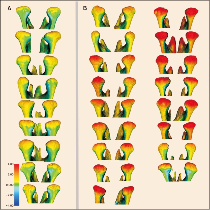
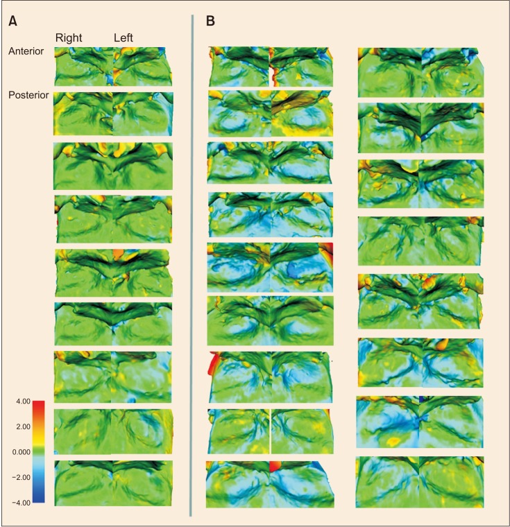
Methods: Seventeen patients with Class II division 1 malocclusion treated using Twin-block and nine untreated patients with a similar malocclusion were included in this research. We collected their cone beam computed tomography (CBCT) data from before and 8 months after treatment. Segmentations were constructed using ITK-SNAP. Condylar volume and superficial area were measured using 3D Slicer. The 3D landmarks were identified on CBCT images by using Dolphin software to assess the condylar positional relationship. 3D models of the mandible and glenoid fossa of the patients were constructed and registered via voxel-based superimposition using 3D Slicer. Thereafter, skeletal changes could be visualized using 3DMeshMetric in any direction of the superimposition on a color-coded map. All the superimpositions were measured using the same scale on the distance color-coded map, in which red color represents overgrowth and blue color represents resorption.
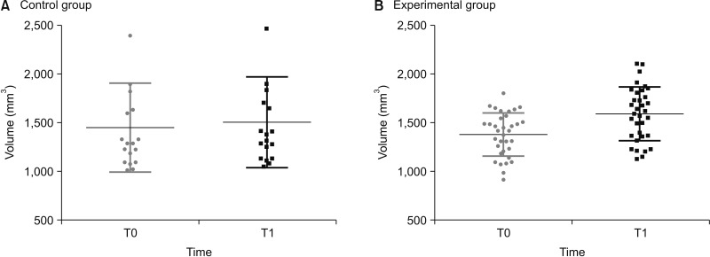
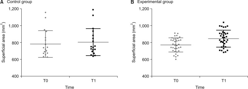
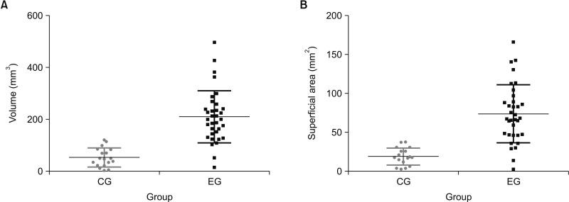
Results: Significant differences were observed in condylar volume, superficial area, and condylar position in both groups after 8 months. Compared with the control group (CG), the Twin-block group exhibited more obvious condyle-fossa modifications and joint positional changes. Moreover, on the color-coded map, more obvious condyle-fossa modifications could be observed in the posterior and superior directions in the Twin-block group than in the CG.
Conclusions: We successfully established a 3D method for measuring and evaluating TMJ changes caused by Twin-block treatment. The treatment produced a larger condylar size and caused condylar positional changes.
Keywords: Computed tomography; Functional; Temporomandibular joint; Three-dimensional cephalometrics.
© 2020 The Korean Association of Orthodontists.
Conflict of interest statement
CONFLICTS OF INTEREST: No potential conflict of interest relevant to this article was reported.
Figures
References
-
- Baccetti T, Franchi L, Toth LR, McNamara JA., Jr Treatment timing for Twin-block therapy. Am J Orthod Dentofacial Orthop. 2000;118:159–170. - PubMed
-
- Jena AK, Duggal R, Parkash H. Skeletal and dentoalveolar effects of Twin-block and bionator appliances in the treatment of Class II malocclusion: a comparative study. Am J Orthod Dentofacial Orthop. 2006;130:594–602. - PubMed
-
- Lund DI, Sandler PJ. The effects of Twin Blocks: a prospective controlled study. Am J Orthod Dentofacial Orthop. 1998;113:104–110. - PubMed
-
- Morris DO, Illing HM, Lee RT. A prospective evaluation of Bass, Bionator and Twin Block appliances. Part II--The soft tissues. Eur J Orthod. 1998;20:663–664. - PubMed
-
- Trenouth MJ. Cephalometric evaluation of the Twin-block appliance in the treatment of Class II Division 1 malocclusion with matched normative growth data. Am J Orthod Dentofacial Orthop. 2000;117:54–59. - PubMed
LinkOut - more resources
Full Text Sources