Progesterone Prolongs Viability and Anti-inflammatory Functions of Explanted Preterm Ovine Amniotic Membrane
- PMID: 32258004
- PMCID: PMC7089934
- DOI: 10.3389/fbioe.2020.00135
Progesterone Prolongs Viability and Anti-inflammatory Functions of Explanted Preterm Ovine Amniotic Membrane
Abstract
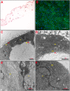
Amniotic membrane (AM) is considered an important medical device with many applications in regenerative medicine. The therapeutic properties of AM are due to its resistant extracellular matrix and to the large number of bioactive molecules released by its cells. An important goal that still remains to be achieved is the identification of cultural and preservation protocols able to maintain in time the membrane morphology and the biological properties of its cells. Recently, our research group demonstrated that progesterone (P4) is crucial in preventing the loss of the epithelial phenotype of amniotic epithelial cells in vitro. Followed by this premise, it has been evaluated whether P4 may also affect AM properties in a short-term culture. Results confirm that P4 preserves AM integrity and architecture with respect to untreated AM, which showed alterations in morphology. Transmission electron microscopy (TEM) analyses demonstrate that P4 also maintains unaltered cell-cell junctions, nuclear status, and intracellular organelles. On the contrary, an untreated AM experienced an extensive cell death and a strong reduction of immunomodulatory properties, measured in terms of anti-inflammatory cytokine expression and secretion. Overall, these results could open to new strategies to ameliorate the protocols for cryopreservation and tissue culture, which represent preliminary stages of AM application in regenerative medicine.
Keywords: amniotic epithelial stem cells; amniotic membrane; immunomodulation; progesterone; regenerative medicine; tissue culture.
Copyright © 2020 Canciello, Teti, Mazzotti, Falconi, Russo, Giordano and Barboni.
Figures


Similar articles
-
Amniotic Membrane and Amniotic Epithelial Cell Culture.Methods Mol Biol. 2024;2749:135-149. doi: 10.1007/978-1-0716-3609-1_13. Methods Mol Biol. 2024. PMID: 38133781
-
Optimizing amniotic membrane tissue banking protocols for ophthalmic use.Cell Tissue Bank. 2016 Sep;17(3):387-97. doi: 10.1007/s10561-016-9568-3. Epub 2016 Jul 18. Cell Tissue Bank. 2016. PMID: 27430235
-
Progesterone prevents epithelial-mesenchymal transition of ovine amniotic epithelial cells and enhances their immunomodulatory properties.Sci Rep. 2017 Jun 19;7(1):3761. doi: 10.1038/s41598-017-03908-1. Sci Rep. 2017. PMID: 28630448 Free PMC article.
-
How preparation and preservation procedures affect the properties of amniotic membrane? How safe are the procedures?Burns. 2020 Sep;46(6):1254-1271. doi: 10.1016/j.burns.2019.07.005. Epub 2019 Aug 21. Burns. 2020. PMID: 31445711 Review.
-
An important detail that is still not clear in amniotic membrane applications: How do we store the amniotic membrane best?Cell Tissue Bank. 2024 Mar;25(1):339-347. doi: 10.1007/s10561-023-10121-0. Epub 2024 Jan 9. Cell Tissue Bank. 2024. PMID: 38191687 Review.
Cited by
-
Unveiling the immunomodulatory shift: Epithelial-mesenchymal transition Alters immune mechanisms of amniotic epithelial cells.iScience. 2023 Aug 9;26(9):107582. doi: 10.1016/j.isci.2023.107582. eCollection 2023 Sep 15. iScience. 2023. PMID: 37680464 Free PMC article.
-
Amniotic Epithelial Stem Cells Counteract Acidic Degradation By-Products of Electrospun PLGA Scaffold by Improving Their Immunomodulatory Profile In Vitro.Cells. 2021 Nov 18;10(11):3221. doi: 10.3390/cells10113221. Cells. 2021. PMID: 34831443 Free PMC article.
-
Graphene oxide accelerates TGFβ-mediated epithelial-mesenchymal transition and stimulates pro-inflammatory immune response in amniotic epithelial cells.Mater Today Bio. 2023 Aug 2;22:100758. doi: 10.1016/j.mtbio.2023.100758. eCollection 2023 Oct. Mater Today Bio. 2023. PMID: 37600353 Free PMC article.
-
Amphiregulin orchestrates the paracrine immune-suppressive function of amniotic-derived cells through its interplay with COX-2/PGE2/EP4 axis.iScience. 2024 Jul 14;27(8):110508. doi: 10.1016/j.isci.2024.110508. eCollection 2024 Aug 16. iScience. 2024. PMID: 39156643 Free PMC article.
-
Amniotic membrane, a novel bioscaffold in cardiac diseases: from mechanism to applications.Front Bioeng Biotechnol. 2024 Dec 20;12:1521462. doi: 10.3389/fbioe.2024.1521462. eCollection 2024. Front Bioeng Biotechnol. 2024. PMID: 39758951 Free PMC article. Review.
References
-
- Abdulkareem T. A., Eidan S. M., Ishak M. A., Al-Sharifi S. A., Alnimer M. A., Passavant C. W., et al. . (2012). Pregnancy-specific protein B (PSPB), progesterone and some biochemical attributes concentrations in the fetal fluids and serum and its relationship with fetal and placental characteristics of Iraqi riverine buffalo (Bubalus bubalis). Anim. Reprod. Sci. 130, 33–41. 10.1016/j.anireprosci.2012.01.002 - DOI - PubMed
-
- Barboni B., Mangano C., Valbonetti L., Marruchella G., Berardinelli P., Martelli A., et al. . (2013). Synthetic bone substitute engineered with amniotic epithelial cells enhances bone regeneration after maxillary sinus augmentation. PLoS ONE 8:e63256. 10.1371/journal.pone.0063256 - DOI - PMC - PubMed
LinkOut - more resources
Full Text Sources

