

Mucoepidermoid Carcinoma of the Lacrimal Sac: Clinical-Pathologic Analysis, Including Molecular Genetics
- PMID: 32258022
- PMCID: PMC7109434
- DOI: 10.1159/000502699
Mucoepidermoid Carcinoma of the Lacrimal Sac: Clinical-Pathologic Analysis, Including Molecular Genetics
Abstract
Purpose: The aim of this study was to assess whether mucoepidermoid carcinoma of the lacrimal sac is a counterpart of CRTC1/3-MAML2 gene fusion-related salivary gland mucoepidermoid carcinoma.
Methods: In this retrospective observational case series, pathology records were searched for all cases of lacrimal sac mucoepidermoid carcinoma diagnosed between 1990 and 2018. Data collected included demographics, clinical findings, management, and follow-up. Pathologic parameters assessed included tumor morphology, immunohistochemistry, and MAML2 and EGFR fluorescence in situ hybridization (FISH) studies.
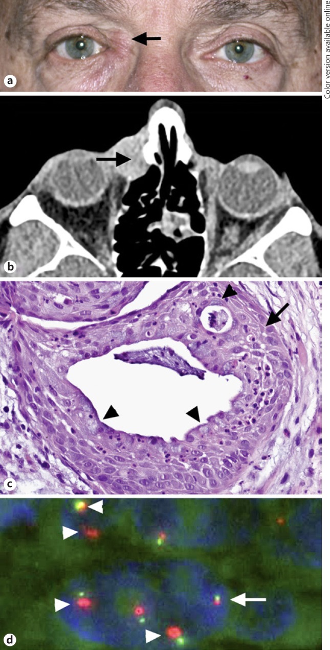
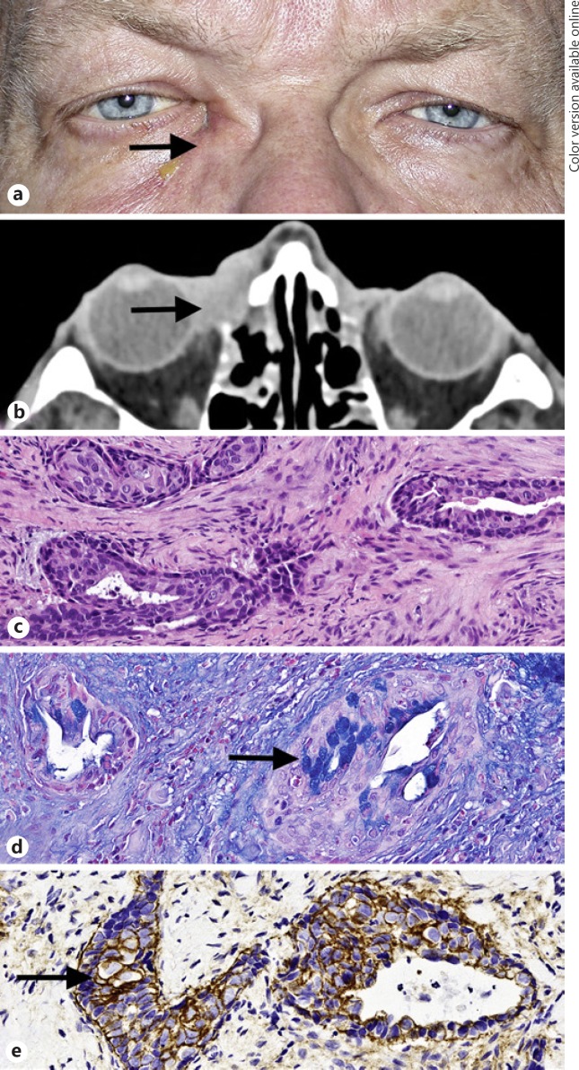
Results: Six patients with mucoepidermoid carcinoma of the lacrimal sac, 5 males and 1 female, with a median age of 63 years (range 24-66) were identified. Five tumors were managed with radical resection and 1 patient underwent orbital exenteration. None of the patients developed recurrence or metastases with an average follow-up of 18 months (range 13-23). All tumors had morphologic and immunohistochemical features of mucoepidermoid carcinoma and overexpressed EGFR. MAML2 FISH was negative for MAML2 rearrangement in all tumors. EGFR FISH demonstrated EGFR amplification in 1 tumor.
Conclusions: Mucoepidermoid carcinoma of the lacrimal sac is not a lacrimal sac counterpart of CRTC1/3-MAML2 gene fusion-related salivary gland mucoepidermoid carcinoma. EGFR pathway activation and EGFR amplification in a subset of these neoplasms suggest the potential role for anti-EGFR agents.
Keywords: Adenosquamous carcinoma; CRTC1/3-MAML2; EGFR; Lacrimal sac carcinoma; Lacrimal sac mucoepidermoid carcinoma; MAML2; Mucoepidermoid carcinoma.
Copyright © 2019 by S. Karger AG, Basel.
Conflict of interest statement
The authors have no conflicts of interest to declare. None of the authors have relevant financial relationships with commercial interests.
Figures
References
-
- Stefanyszyn MA, Hidayat AA, Pe'er JJ, Flanagan JC. Lacrimal sac tumors. Ophthal Plast Reconstr Surg. 1994 Sep;10((3)):169–84. - PubMed
-
- Fliss DM, Freeman JL, Hurwitz JJ, Heathcote JG. Mucoepidermoid carcinoma of the lacrimal sac: a report of two cases, with observations on the histogenesis. Can J Ophthalmol. 1993 Aug;28((5)):228–35. - PubMed
-
- Lee SB, Kim KN, Lee SR, Bernardino CR. Mucoepidermoid carcinoma of the lacrimal sac after dacryocystectomy for squamous papilloma. Ophthal Plast Reconstr Surg. 2011 Mar-Apr;27((2)):e44–6. - PubMed
-
- Brar ST, Meyer D. Diagnosis and management of mucoepidermoid carcinoma of the lacrimal duct. Orbit. 2011 Jan;30((1)):34–6. - PubMed
-
- Yuksel D, Kosker M, Saribas F, Simsek S. Surgical treatment of mucoepidermoid carcinoma of the lacrimal sac. Semin Ophthalmol. 2014 Mar;29((2)):70–2. - PubMed
LinkOut - more resources
Full Text Sources
Research Materials
Miscellaneous
