. 2020 Mar;6(2):145-150.
doi: 10.1159/000501295. Epub 2019 Aug 9.

Massive Silicone-Induced Orbital Granuloma

Affiliations

Massive Silicone-Induced Orbital Granuloma

Ann Shue et al. Ocul Oncol Pathol. 2020 Mar.

Abstract

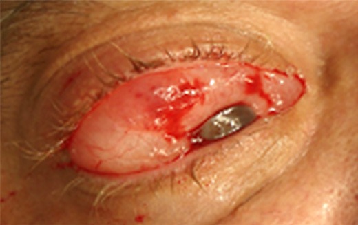
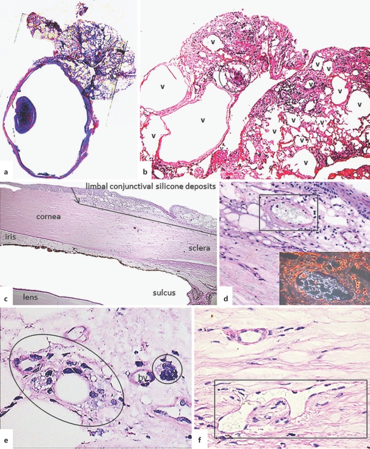
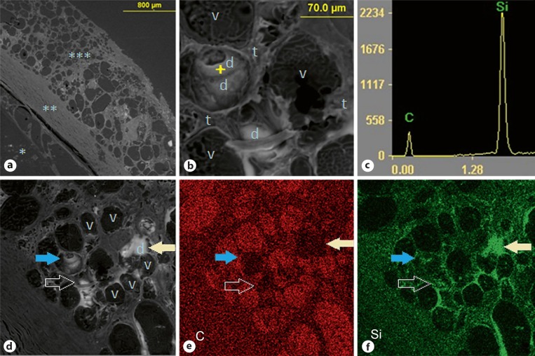
We report a large subconjunctival-orbital granuloma in a 51-year-old male presenting with a blind painful right eye and marked chemosis 15 months after undergoing vitrectomy and silicone oil retinal tamponade for retinal detachment with no reported intraoperative complications. Gross and histopathologic examination of the enucleated eye and episcleral tumor revealed a bosselated mass measuring 17 × 10 × 5 mm containing prominent vacuoles with surrounding epithelioid histiocytes and foreign body multinucleated giant cells. Such a large silicone-induced orbital granuloma following uncomplicated retinal surgery in a grossly intact eye has not been previously reported to the authors' knowledge. High intraocular pressure and emulsification of oil may facilitate silicone extravasation through scleral wounds after retinal surgery.

Keywords: Case report; Scanning electron microscope-energy dispersive spectroscopy; Silicone-induced orbital granuloma.


Conflict of interest statement

The authors have no conflicts of interest to declare.

Figures

Fig. 1
Fig. 1
Clinical presentation. The patient presented with a large mass in the right superior lateral orbit 15 months after right eye retinal detachment repair with silicone oil tamponade. Abduction and supraduction of the blind and painful eye were severely restricted.
Fig. 2
Fig. 2
Histopathologic features of the enucleated eye showing extra- and intraocular granulomatous inflammation due to silicone. a Relationship of orbital mass adherent to intact sclera. Periodic Acid-Schiff, whole slide image. b Orbital tissue with granulomatous reaction (circled) to silicone deposits in the variably sized vacuoles (v), now empty due to processing. Hematoxylin-eosin. Original magnification, ×125. c–f Intraocular tissue deposits of silicone. c Silicone deposits in the limbal conjunctiva as well as angle closure (other structures are labeled for orientation). Periodic Acid-Schiff. Original magnification, ×40. d Subepithelial conjunctiva shows retained refractile silicone in several vacuoles shown best by dark-field illumination (inset). Periodic Acid-Schiff. Original magnification, ×200. e Silicone phagocytosed by macrophage giant cells (circled). bv, blood vessel. Periodic Acid-Schiff. ×400. f Refractile silicone in Schlemm's canal (in rectangle). Periodic Acid-Schiff. ×400.
Fig. 3
Fig. 3
SEM-EDS images (backscattered electron images; brightness depends on average atomic number of tissue). a Low-power section, labeled * vitreous cavity, ** sclera, *** limbal-episcleral nodule, or an extension of a larger mass, with variably sized vacuoles. b A high-power section of the limbal-episcleral nodule showing scattered deposits, one of which (indicated by the “+” sign) was analyzed by EDS. The tissue components are labeled as follows: v, vacuoles (mostly empty); t, intervacuolar tissue; d, deposits. c EDS spectrum of the deposit indicated in b, showing a prominent silicon (Si) peak relative to the carbon (C) peak from the paraffin substrate. d Lower magnification of the area shown in b that reveals further deposits (d, brighter white) as well as empty vacuoles (v). e, f EDS distribution maps for carbon (C, shown in red) and silicon (Si, shown in green), which confirm the spatial distribution of silicon in (brighter) deposits and to a lesser degree in intervacuolar tissue, and absence in empty vacuoles, where carbon from the paraffin substrate is the major finding. The matching arrows point to corresponding areas in images d–f.

References

    1. Abel AD, Carlson JA, Bakri S, Meyer DR. Sclerosing lipogranuloma of the orbit after periocular steroid injection. Ophthalmology. 2003 Sep;110((9)):1841–5. - PubMed
    1. Davies GB, Wong PL. Sclerosing lipo-granuloma in the orbit. Br J Ophthalmol. 1958 Nov;42((11)):697–701. - PMC - PubMed
    1. Couch SM, Harocopos GJ, Holds JB. Orbital silicone oil granuloma discovered during enucleation. Arch Ophthalmol. 2012 Aug;130((8)):1083–5. - PubMed
    1. Lee JH, Kim YD, Woo KI, Kong M. Subconjunctival and orbital silicone oil granuloma (siliconoma) complicating intravitreal silicone oil tamponade. Case Rep Ophthalmol Med. 2014;2014:686973. - PMC - PubMed
    1. Travis WD, Balogh K, Abraham JL. Silicone granulomas. A review of the literature and report of 3 cases. Hum Pathol. 1985;16:19–27. - PubMed