Case Reports
. 2020 Mar 13:2020:8797493.
doi: 10.1155/2020/8797493. eCollection 2020.

Biomechanical Analysis of Various Reconstructive Methods for the Mandibular Body and Ramus Defect Using a Free Vascularized Fibula Flap

Affiliations
Case Reports

Biomechanical Analysis of Various Reconstructive Methods for the Mandibular Body and Ramus Defect Using a Free Vascularized Fibula Flap

Xian Li et al. Biomed Res Int. .

Abstract

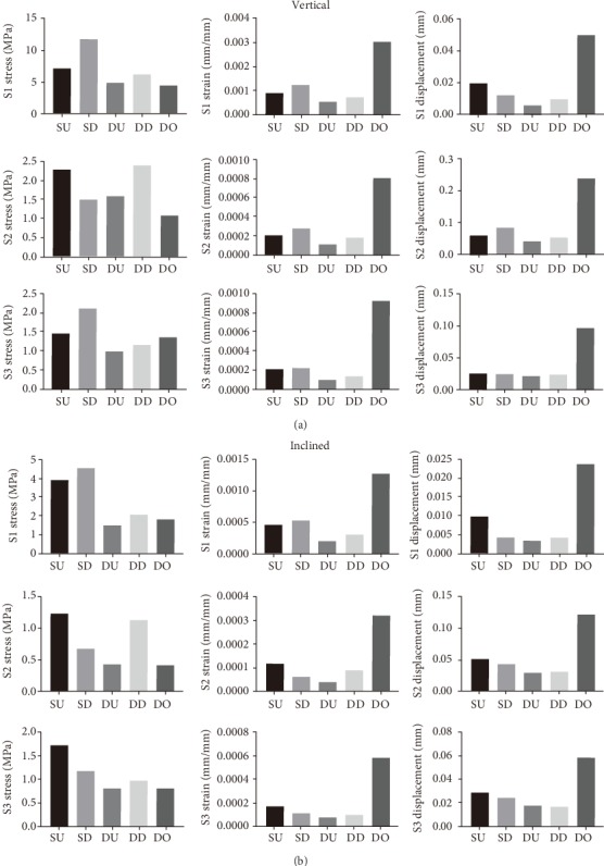
Several different methods exist for reconstructing the mandibular body and ramus defect with the use of a free vascularized fibula flap, but none have adequately addressed the long-term mechanical stability and osseointegration. The aim of this study is to compare the biomechanics of different surgical methods and to investigate the best approach for reconstructing the mandibular body and ramus defect. Five finite element models based on different reconstructive methods were simulated. Stress, strain, and displacement of connective bone sections were calculated for five models and compared. The models were printed using a 3D printer, and stiffness was measured using an electromechanical universal testing machine. The postoperative follow-up cone beam computed tomography (CBCT) was taken at different time points to analyze bone mineral density of connective bone sections. The results showed that the "double up" (DU) model was the most efficient for reconstructing a mandibular body and ramus defect by comparing the mechanical distribution of three sections under vertical and inclined loading conditions of 100 N. The stiffness detection showed that stiffness in the DU and "double down" (DD) models was higher compared with the "single up" (SU), "single down" (SD), and "distraction osteogenesis" (DO) models. We used the DU model for the surgery, and postoperative follow-up CBCT showed that bone mineral density of each fibular connective section increased gradually with time, plateauing at 12 weeks. We conclude that a free vascularized fibula flap of the DU type was the best approach for the reconstruction of the mandibular body and ramus defect. Preoperative finite element analysis and stiffness testing were shown to be very useful for maxillofacial reconstruction.


Conflict of interest statement

None of the authors has a proprietary or financial interest in any product mentioned.

Figures

Figure 1
Figure 1
FE models and the number of nodes and elements of FE models.
Figure 2
Figure 2
Each model was applied with a vertical and inclined force of 100 N each. The three bone connective sections (S1, S2, and S3) were marked by the golden boxes and amplified.
Figure 3
Figure 3
(a, c) The mechanical distribution in S1 in the SU, SD, DU, DD, and DO models, under vertical and inclined loading conditions of 100 N, respectively. (b, d) Histograms comparing the maximum values of stress, strain, and displacement in the five models under vertical and inclined loading conditions of 100 N, respectively.
Figure 4
Figure 4
(a, c) The mechanical distribution in S2 in the SU, SD, DU, DD, and DO models, under vertical and inclined loading conditions of 100 N each. (b, d) Histograms comparing the maximum values of stress, strain, and displacement in S2 in the five models under vertical and inclined loading conditions of 100 N, respectively.
Figure 5
Figure 5
(a, c) The mechanical distribution in S3 in the SU, SD, DU, DD, and DO models under vertical and inclined loading conditions of 100 N, respectively. (b, d) Histograms comparing the maximum values of stress, strain, and displacement in S3 in the five models under vertical and inclined loading conditions of 100 N, respectively.
Figure 6
Figure 6
Medians of vertical and inclined forces in S1, S2, and S3. (a) Medians of stress, strain, and displacement values of S1, S2, and S3 in the SU, SD, DU, DD, and DO models under vertical loading conditions of 100 N. (b) Medians of stress, strain, displacement values of S1, S2, and S3 in the SU, SD, DU, DD, and DO models under inclined loading conditions of 100 N.
Figure 7
Figure 7
Stiffness detection and statistical analysis of all models. (a) Five reconstructive models were printed out, and stiffness was measured using an electromechanical universal testing machine. (b) Scatterplot comparison of stiffness measurements. Statistical analysis: one-way ANOVA (p < 0.05, ∗∗p < 0.01, ∗∗∗p < 0.005, ∗∗∗∗p < 0.001).
Figure 8
Figure 8
Digital design and operation. (a) Digital design of mandibular tumor resection. (b) 3D printed osteotomy guided plates of the mandible. (c) Intraoperative resection of mandibular tumor resection. (d) Digital design of fibular resection. (e) 3D printed osteotomy guided plate of the fibula. (f) Intraoperative resection of fibular resection. (g) Digital design of fibular segments' emplacement. (h) 3D printed fibular emplacement guided plates. (i) Postoperative CBCT of fibular reconstruction.
Figure 9
Figure 9
Postoperative CBCT and bone mineral density statistics in connecting sections. (a–c) Postoperative follow-up CBCT at 2w, 12w, and 24w. (d–f) Histogram comparison of bone mineral density of each fibular connecting section (S1, S2, and S3) at 2w, 12w, and 24w. Statistical analysis: one-way ANOVA (p < 0.05, ∗∗p < 0.01, ∗∗∗p < 0.005, ∗∗∗∗p < 0.001).

Similar articles

Cited by

References

    1. Hidalgo D. A., Rekow A. A review of 60 consecutive fibula free flap mandible reconstructions. Plastic and Reconstructive Surgery. 1995;96(3):585–596. doi: 10.1097/00006534-199509000-00010. - DOI - PubMed
    1. Vittayakittipong P., Jarudejkajon J., Kirirat P., Chaijaroonkhanarak W., Chaisiwamongkol K. Feasibility of the vascularized fibula bone graft for reconstruction of the mandible: a cadaveric study. International Journal of Oral and Maxillofacial Surgery. 2016;45(8):960–963. doi: 10.1016/j.ijom.2015.11.016. - DOI - PubMed
    1. Shroff S. S., Nair S. C., Shah A., Kumar B. Versatility of fibula free flap in reconstruction of facial defects: a center study. Journal of Maxillofacial and Oral Surgery. 2017;16(1):101–107. doi: 10.1007/s12663-016-0930-6. - DOI - PMC - PubMed
    1. Inbal A., Gur E., Zaretski A., Barnea Y., Khafif A., Amir A. The "origami" composite free fibula flap for complex defects of the mandible, floor of the mouth, and tongue. Journal of Oral and Maxillofacial Surgery. 2015;73(8):1617–1626. doi: 10.1016/j.joms.2015.02.004. - DOI - PubMed
    1. Shokri T., Stahl L. E., Kanekar S. G., Goyal N. Osseous changes over time in free fibular flap reconstruction. The Laryngoscope. 2019;129(5):1113–1116. doi: 10.1002/lary.27337. - DOI - PubMed

Publication types