. 2019 Sep 9;5(9):4551-4563.
doi: 10.1021/acsbiomaterials.9b00505. Epub 2019 Aug 1.

A Visible Light-Cross-Linkable, Fibrin-Gelatin-Based Bioprinted Construct with Human Cardiomyocytes and Fibroblasts

Affiliations

A Visible Light-Cross-Linkable, Fibrin-Gelatin-Based Bioprinted Construct with Human Cardiomyocytes and Fibroblasts

Shweta Anil Kumar et al. ACS Biomater Sci Eng. .

Abstract

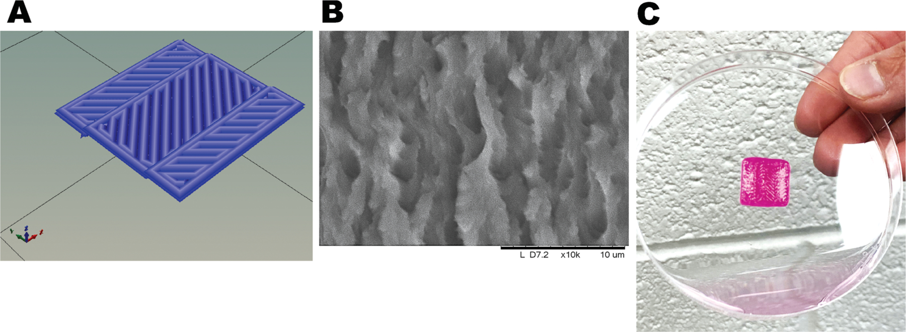
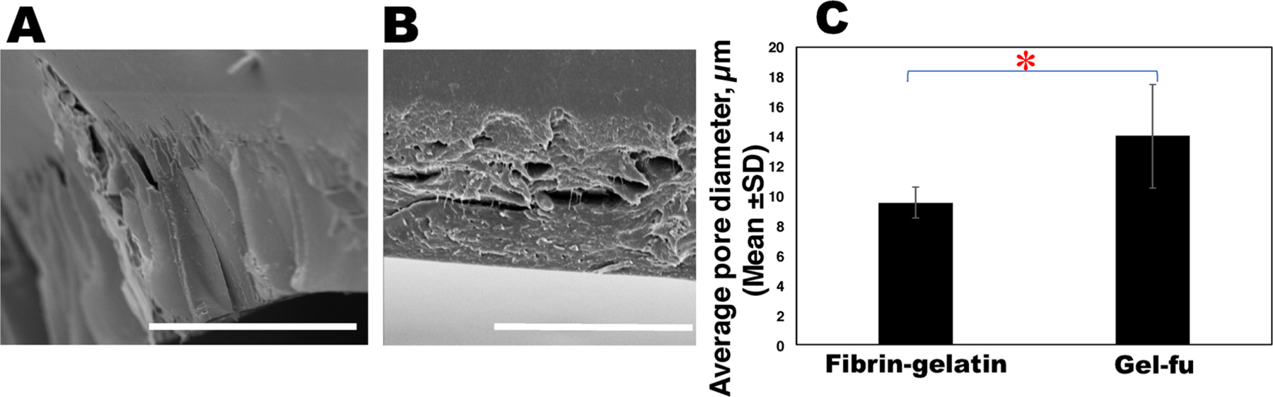
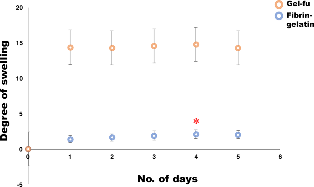
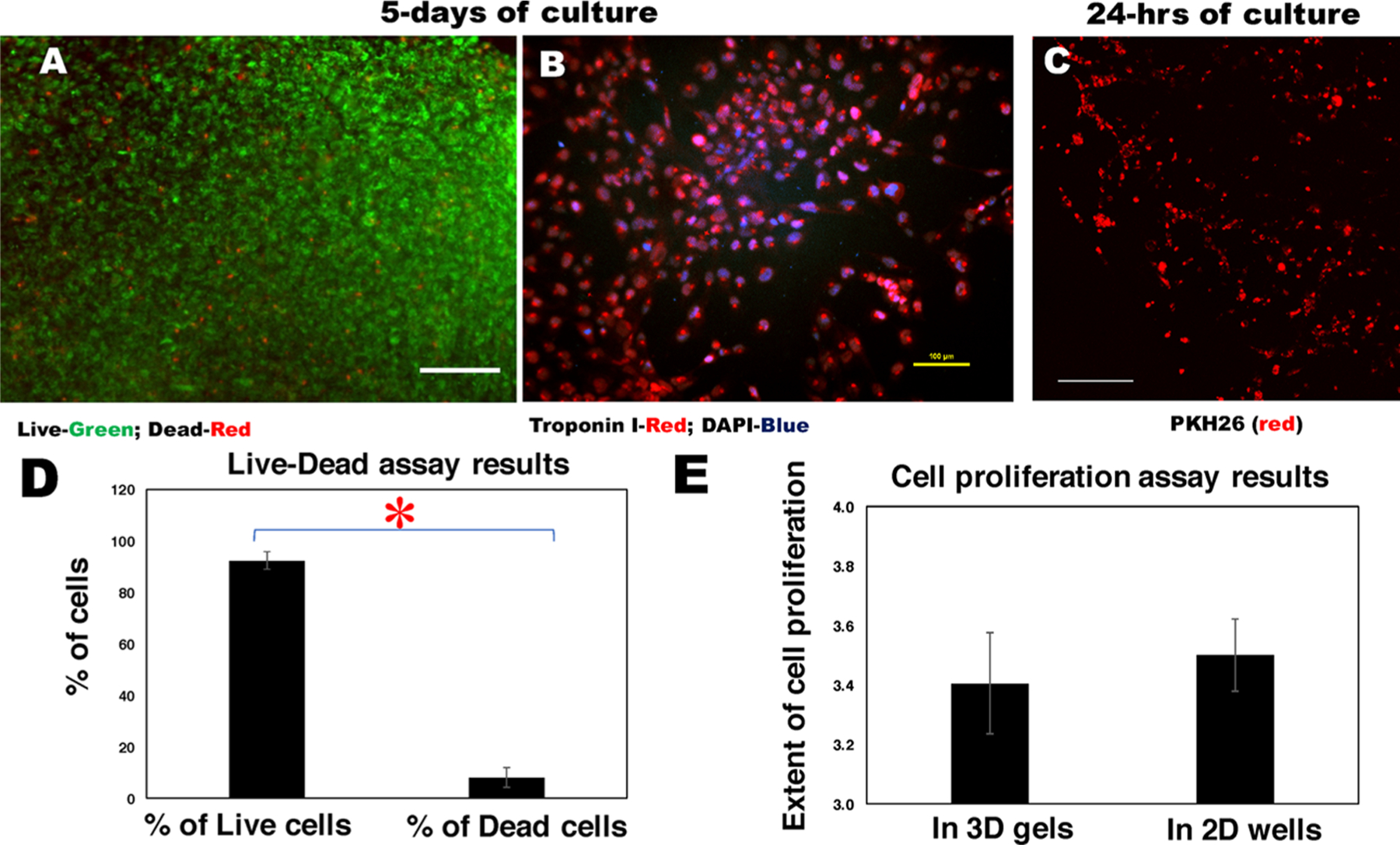
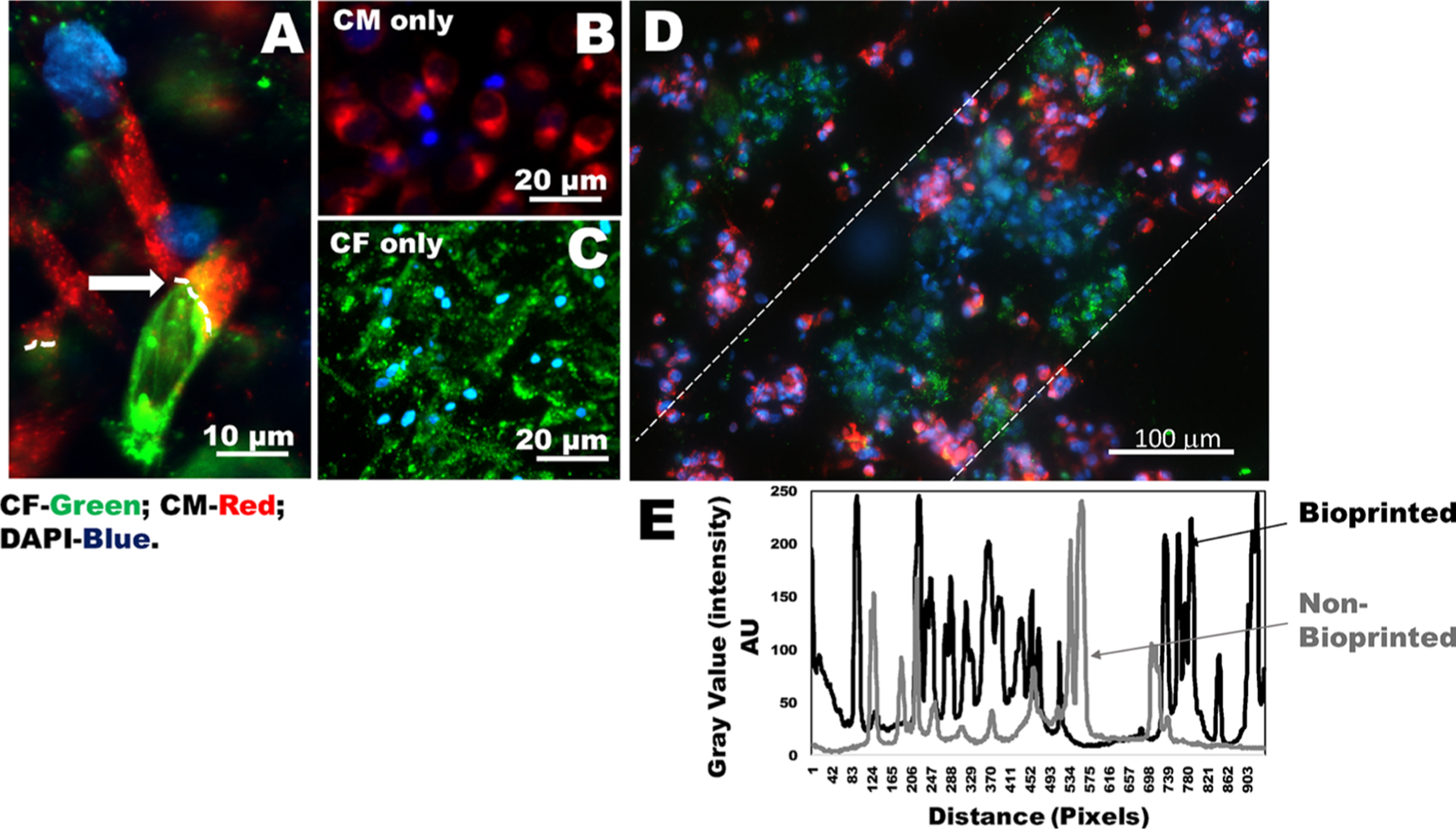
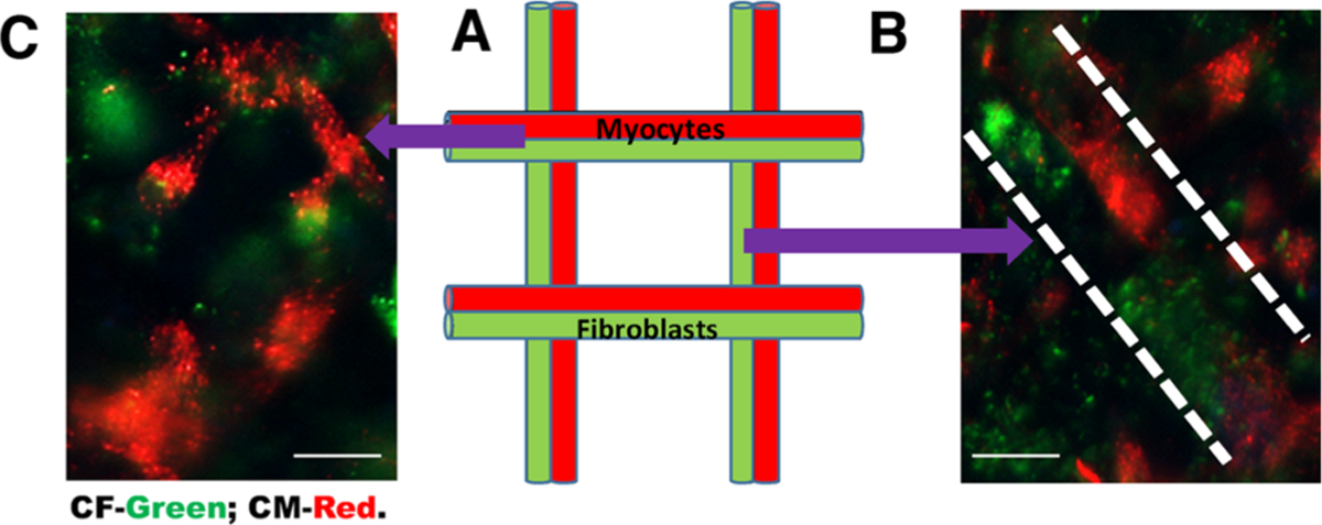
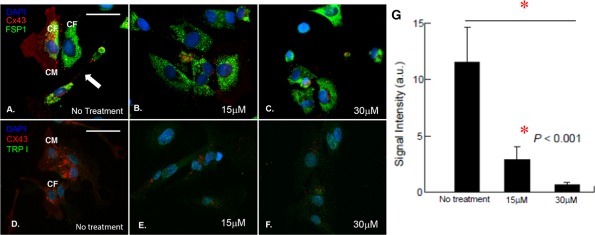
In this study, fibrin was added to a photo-polymerizable gelatin-based bioink mixture to fabricate cardiac cell-laden constructs seeded with human induced pluripotent stem cell-derived cardiomyocytes (iPS-CM) or CM cell lines with cardiac fibroblasts (CF). The extensive use of platelet-rich fibrin, its capacity to offer patient specificity, and the similarity in composition to surgical glue prompted us to include fibrin in the existing bioink composition. The cell-laden bioprinted constructs were cross-linked to retain a herringbone pattern via a two-step procedure including the visible light cross-linking of furfuryl-gelatin followed by the chemical cross-linking of fibrinogen via thrombin and calcium chloride. The printed constructs revealed an extremely porous, networked structure that afforded long-term in vitro stability. Cardiomyocytes printed within the sheet structure showed excellent viability, proliferation, and expression of the troponin I cardiac marker. We extended the utility of this fibrin-gelatin bioink toward coculturing and coupling of CM and cardiac fibroblasts (CF), the interaction of which is extremely important for maintenance of normal physiology of the cardiac wall in vivo. This enhanced "cardiac construct" can be used for drug cytotoxicity screening or unraveling triggers for heart diseases in vitro.

Keywords: 3D bioprinting; biofabrication; cardiac tissue; fibrinogen; furfuryl–gelatin; thrombin.


Conflict of interest statement

The authors declare no competing financial interest.

Figures

Figure 1.
Figure 1.
Gross morphology of the structure printed using a fibrin–gelatin gel. (A) .stl file image. (B) Representative SEM en face image of a characteristic 3D printed pattern. (C) Herringbone construct casted with the bioprinter.
Figure 2.
Figure 2.
SEM analysis for pore size estimation. (A) Representative SEM image of the edge of a characteristic fibrin–gelatin film. At least five representative images were acquired per sample and used to determine the average pore size depicted in (C), in comparison with the gel-fu (f-gelatin) structures as depicted in (B). (C) Plot comparing the average pore diameters for the fibrin–gelatin (current study)- and f-gelatin (gel-fu; previous study)-based constructs. The average pore diameters of the fibrin–gelatin films were significantly reduced (*P < 0.05) in comparison with gel-fu patterns from our previous study. In (A), the scale bar corresponds to 50 μm, and in (B), it corresponds to 20 μm.
Figure 3.
Figure 3.
Swelling analysis. Degree of swelling (mean ± SD) for a characteristic fibrin–gelatin-based pattern (blue circles) for over a period of 5 days for which the maximum degree of swelling was attained at day 4, significantly greater (*P < 0.05) than what was seen at previous time points. Beyond day 4, until day 5, the degree of swelling apparently reached equilibrium as these values did not seem to bear any statistical differences when analyzed. For comparison, a characteristic swelling degradation curve from a construct made with gel-fu (orange circles), as reported in our previous study, is also included as controls.
Figure 4.
Figure 4.
Rheology analysis of fibrin–gelatin-based square structures. (A) Representative storage and loss moduli and complex viscosity from one characteristic gel sample. Therefore, it does not have error bars. (B) Average storage and loss moduli for similar sample patterns (#1 and #2, n = 2) analyzed during rheological characterization.
Figure 5.
Figure 5.
Cell retention, viability, and proliferation. Characteristic images of human CM cell lines that were bioprinted within fibrin–gelatin patterns and (A) imaged using Live (green)/Dead (red) assay and (B) stained with troponin I antibody (red) and DAPI (blue) after 5 days of culture. In (A), the scale bar depicts 200 μm, and in (B), it is 100 μm. (C) PKH26 (red)-prestained cells bioprinted using a herringbone design were seen to align along this pattern after 24 h of culture in a maximum intensity projection of a Z-stack of confocal microscope images. Scale bar depicts 250 μm. (D) Quantification of cell viability estimated by Live/Dead assay and (E) the extent of live cell proliferation in samples. Statistically significant differences are denoted by an asterisk (*P < 0.05).
Figure 6.
Figure 6.
Heterocellular coupling of CM and CF. (A) Human CM (prestained using PKH26, red) and CF (prestained using PKH67, green) coupled in 2D wells in the ratio of CM/CF (1:1) shown by white arrows and dotted lines. (B, C) Images of controls for either CM or CF (in 2D wells). (D) Results of heterocellular coupling between prestained CM (red) and CF (green) from 3D bioprinted constructs. Nonprinted controls that included cells mixed in gels are shown in Figure S5. The white dashed lines in (D) denote that the cells are confined to their printed patterns. However, these samples were fixed prior to DAPI staining, and the cells were not viable during imaging. To confirm heterocellular coupling using viable cells, we report results in Figure 7. (E) Intensity values within the white dotted lines in a bioprinted sample (black line) versus a nonbioprinted sample (gray line). The average intensity values for the bioprinted samples exhibited more frequent peaks with respect to distance, depicting a higher frequency of cells present in the preferred orientation compared to samples that were nonbioprinted.
Figure 7.
Figure 7.
Heterocellular coupling of nonfixed CM and CF in 3D bioprinted gels. Viable and nonfixed cells used to confirm heterocellular coupling between CM and CF. (A) Schematic of a lattice grid pattern for coculturing of CM (red) and CF (green). (B) Human CM (red) and CF (green) coupled when confined to two adjacent line patterns (as denoted by white dashed lines). (C) Representative cell coupling in junctions of the grid pattern. Although cell coupling may be occurring in cells mixed and cross-linked within gels, it is very difficult to confirm in samples, which are not patterned distinctively using bioprinting, as shown in Figure S5. Scale bars depict 10 μm.
Figure 8.
Figure 8.
Confirmation of heterocellular coupling between CM and CF. (A–C) Cocultured cells immunostained with Cx43 (red), FSP-1 (green), and DAPI (blue) with no Gap26 treatment (A) and with 15 μM (B) and 30 μM Gap26 (C). (D–F) Cocultured cells immunostained with Cx43 (red), troponin I (TRP I, green), and DAPI (blue) with no Gap26 treatment (D) and with 15 μM (E) and 30 μM Gap26 (F). Scale bars in (A)–(F) are of the same size and correspond to 50 μM. (G) Average signal intensity of Cx43 expression in cultures with no Gap26 treatment and with 15 μM (B) and 30 μM Gap26.

Similar articles

Cited by

References

    1. Asaria P; Elliott P; Douglass M; Obermeyer Z; Soljak M; Majeed A; Ezzati M Acute myocardial infarction hospital admissions and deaths in England: a national follow-back and follow-forward record-linkage study. Lancet Public Health 2017, 2, e191–e201. - PMC - PubMed
    1. Stanford W; Thompson BH; Weiss RM Coronary artery calcification: clinical significance and current methods of detection. AJR, Am. J. Roentgenol 1993, 161, 1139–1146. - PubMed
    1. Ahuja P; Sdek P; MacLellan WR Cardiac myocyte cell cycle control in development, disease, and regeneration. Physiol. Rev 2007, 87, 521–544. - PMC - PubMed
    1. Sun Y; Weber KT Infarct scar: a dynamic tissue. Cardiovasc. Res 2000, 46, 250–256. - PubMed
    1. Thygesen K; Uretsky BF Acute ischaemia as a trigger of sudden cardiac death. Eur. Heart J. Suppl 2004, 6, D88–D90.

LinkOut - more resources