

Mitigating cavitation erosion using biomimetic gas-entrapping microtextured surfaces (GEMS)
- PMID: 32258392
- PMCID: PMC7101208
- DOI: 10.1126/sciadv.aax6192
Mitigating cavitation erosion using biomimetic gas-entrapping microtextured surfaces (GEMS)
Abstract
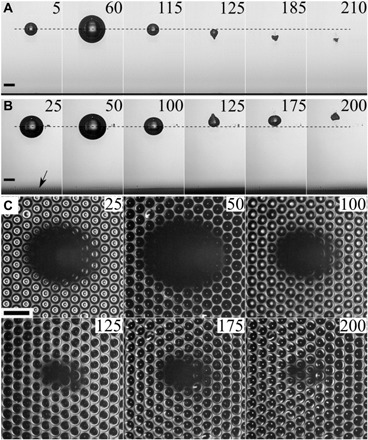
Cavitation refers to the formation and collapse of vapor bubbles near solid boundaries in high-speed flows, such as ship propellers and pumps. During this process, cavitation bubbles focus fluid energy on the solid surface by forming high-speed jets, leading to damage and downtime of machinery. In response, numerous surface treatments to counteract this effect have been explored, including perfluorinated coatings and surface hardening, but they all succumb to cavitation erosion eventually. Here, we report on biomimetic gas-entrapping microtextured surfaces (GEMS) that robustly entrap air when immersed in water regardless of the wetting nature of the substrate. Crucially, the entrapment of air inside the cavities repels cavitation bubbles away from the surface, thereby preventing cavitation damage. We provide mechanistic insights by treating the system as a potential flow problem of a multi-bubble system. Our findings present a possible avenue for mitigating cavitation erosion through the application of inexpensive and environmentally friendly materials.
Copyright © 2020 The Authors, some rights reserved; exclusive licensee American Association for the Advancement of Science. No claim to original U.S. Government Works. Distributed under a Creative Commons Attribution NonCommercial License 4.0 (CC BY-NC).
Figures


Similar articles
-
The effect of scalable PDMS gas-entrapping microstructures on the dynamics of a single cavitation bubble.Sci Rep. 2022 Nov 27;12(1):20379. doi: 10.1038/s41598-022-24746-w. Sci Rep. 2022. PMID: 36437305 Free PMC article.
-
Experimental study on the mesoscale causes of the influence of viscosity on material erosion in a cavitation field.Ultrason Sonochem. 2019 Dec;59:104699. doi: 10.1016/j.ultsonch.2019.104699. Epub 2019 Jul 17. Ultrason Sonochem. 2019. PMID: 31476699
-
Low-intensity ultrasound induced cavitation and streaming in oxygen-supersaturated water: Role of cavitation bubbles as physical cleaning agents.Ultrason Sonochem. 2019 Apr;52:268-279. doi: 10.1016/j.ultsonch.2018.11.025. Epub 2018 Dec 5. Ultrason Sonochem. 2019. PMID: 30573434
-
Modelling cavitation during drop impact on solid surfaces.Adv Colloid Interface Sci. 2018 Oct;260:46-64. doi: 10.1016/j.cis.2018.08.004. Epub 2018 Aug 25. Adv Colloid Interface Sci. 2018. PMID: 30195460 Review.
-
Research on synergistic erosion by cavitation and sediment: A review.Ultrason Sonochem. 2023 May;95:106399. doi: 10.1016/j.ultsonch.2023.106399. Epub 2023 Apr 5. Ultrason Sonochem. 2023. PMID: 37060709 Free PMC article. Review.
Cited by
-
Omni-Directional Assembly of 2D Single-Crystalline Metal Nanosheets.Adv Mater. 2025 Jun;37(24):e2501632. doi: 10.1002/adma.202501632. Epub 2025 Mar 27. Adv Mater. 2025. PMID: 40145860 Free PMC article.
-
Bubble energy generator.Sci Adv. 2022 Jun 24;8(25):eabo7698. doi: 10.1126/sciadv.abo7698. Epub 2022 Jun 24. Sci Adv. 2022. PMID: 35749507 Free PMC article.
-
Superhydrophobicity and size reduction enabled Halobates (Insecta: Heteroptera, Gerridae) to colonize the open ocean.Sci Rep. 2020 May 8;10(1):7785. doi: 10.1038/s41598-020-64563-7. Sci Rep. 2020. PMID: 32385357 Free PMC article.
-
Onset of cavitation and vapor bubble development over hydrophilic and hydrophobic surfaces.Proc Natl Acad Sci U S A. 2025 Jul 8;122(27):e2503033122. doi: 10.1073/pnas.2503033122. Epub 2025 Jul 1. Proc Natl Acad Sci U S A. 2025. PMID: 40591596
-
The effect of scalable PDMS gas-entrapping microstructures on the dynamics of a single cavitation bubble.Sci Rep. 2022 Nov 27;12(1):20379. doi: 10.1038/s41598-022-24746-w. Sci Rep. 2022. PMID: 36437305 Free PMC article.
References
-
- Blake J. R., Gibson D., Cavitation bubbles near boundaries. Annu. Rev. Fluid Mech. 19, 99–123 (1987).
-
- C. E. Brennen, Cavitation and Bubble Dynamics (Cambridge Univ. Press, 2013).
-
- K.-H. Kim, G. Chahine, J.-P. Franc, A. Karimi, Advanced Experimental and Numerical Techniques for Cavitation Erosion Prediction (Springer, 2014), vol. 106.
-
- Philipp A., Lauterborn W., Cavitation erosion by single laser-produced bubbles. J. Fluid Mech. 361, 75–116 (1998).
-
- Haines J. R., Riemer B. W., Felde D. K., Hunn J. D., Pawel S. J., Tsai C. C., Summary of cavitation erosion investigations for the SNS mercury target. J. Nucl. Mater. 343, 58–69 (2005).
LinkOut - more resources
Full Text Sources
