. 2020 Mar 26:3:48.
doi: 10.1038/s41746-020-0255-1. eCollection 2020.

Siamese neural networks for continuous disease severity evaluation and change detection in medical imaging

Affiliations

Siamese neural networks for continuous disease severity evaluation and change detection in medical imaging

Matthew D Li et al. NPJ Digit Med. .

Abstract

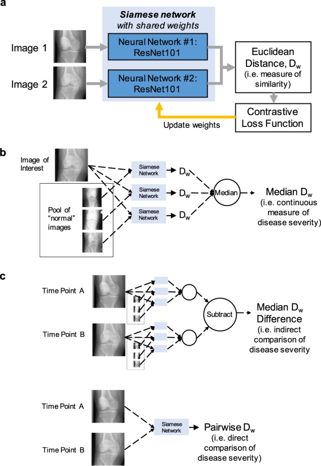
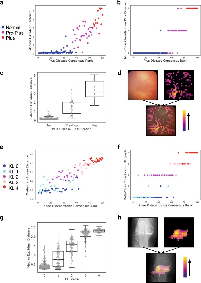
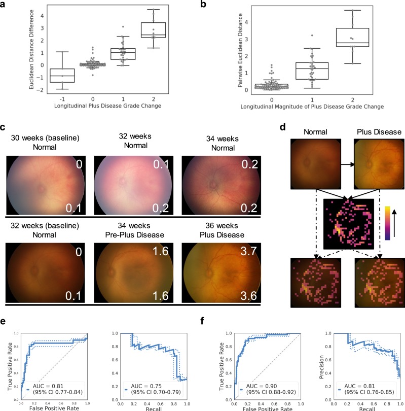
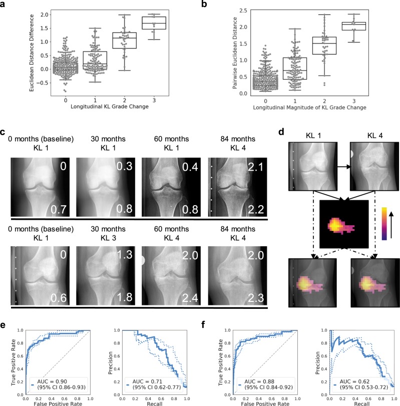
Using medical images to evaluate disease severity and change over time is a routine and important task in clinical decision making. Grading systems are often used, but are unreliable as domain experts disagree on disease severity category thresholds. These discrete categories also do not reflect the underlying continuous spectrum of disease severity. To address these issues, we developed a convolutional Siamese neural network approach to evaluate disease severity at single time points and change between longitudinal patient visits on a continuous spectrum. We demonstrate this in two medical imaging domains: retinopathy of prematurity (ROP) in retinal photographs and osteoarthritis in knee radiographs. Our patient cohorts consist of 4861 images from 870 patients in the Imaging and Informatics in Retinopathy of Prematurity (i-ROP) cohort study and 10,012 images from 3021 patients in the Multicenter Osteoarthritis Study (MOST), both of which feature longitudinal imaging data. Multiple expert clinician raters ranked 100 retinal images and 100 knee radiographs from excluded test sets for severity of ROP and osteoarthritis, respectively. The Siamese neural network output for each image in comparison to a pool of normal reference images correlates with disease severity rank (ρ = 0.87 for ROP and ρ = 0.89 for osteoarthritis), both within and between the clinical grading categories. Thus, this output can represent the continuous spectrum of disease severity at any single time point. The difference in these outputs can be used to show change over time. Alternatively, paired images from the same patient at two time points can be directly compared using the Siamese neural network, resulting in an additional continuous measure of change between images. Importantly, our approach does not require manual localization of the pathology of interest and requires only a binary label for training (same versus different). The location of disease and site of change detected by the algorithm can be visualized using an occlusion sensitivity map-based approach. For a longitudinal binary change detection task, our Siamese neural networks achieve test set receiving operator characteristic area under the curves (AUCs) of up to 0.90 in evaluating ROP or knee osteoarthritis change, depending on the change detection strategy. The overall performance on this binary task is similar compared to a conventional convolutional deep-neural network trained for multi-class classification. Our results demonstrate that convolutional Siamese neural networks can be a powerful tool for evaluating the continuous spectrum of disease severity and change in medical imaging.

Keywords: Diagnosis; Machine learning; Medical imaging; Medical research.


Conflict of interest statement

Competing interestsThe M.F.C is an unpaid member of the scientific advisory board for Clarity Medical Systems (Pleasanton, CA), a consultant for Novartis (Basel, Switzerland), and an initial member of Inteleretina, LLC (Honolulu, HI). J.P.C. and M.F.C. receive research support from Genentech. J.K. is a consultant/advisory board member for Infotech, Soft. The other authors declare no competing interests.

Figures

Fig. 1
Fig. 1. Schematics of Siamese neural network approaches for evaluating disease severity and change on a continuous spectrum.
a Schematic of the Siamese neural network architecture, which takes two images as inputs and outputs the Euclidean distance between the two images (i.e., a measure of similarity). b Schematic of evaluating a single image for disease severity on a continuous spectrum. c Schematic of two approaches for evaluating longitudinal images for disease severity on a continuous spectrum. Dw refers to the Euclidean distance.
Fig. 2
Fig. 2. Siamese neural network outputs can be used to represent a continuous spectrum of disease severity.
a Scatterplot shows the median Euclidean distance versus consensus disease severity rank for 100 retinal photographs ranked by experts for severity of retinopathy of prematurity (plus disease classification). The five retinal photographs with the least severe disease (i.e., most normal) were used as the anchor images to which all the other images were compared. b Scatterplot shows the output of a conventional neural network trained for multi-class classification of plus disease severity. y-axis 0, 1, and 2 indicate ordinal plus disease severity grades, corresponding to normal, pre-plus, and plus disease. c Boxplot* shows the median Euclidean distance calculated in relation to ten randomly sampled “normal” images, separated by plus disease classification. d Illustrative example of an occlusion sensitivity map for visualization of salient areas of the image. e Scatterplot shows the median Euclidean distance versus consensus disease severity rank for 100 knee radiographs ranked by experts for severity of knee osteoarthritis (KL grade). The five knee radiographs with the least severe disease (i.e., most normal) were used as the anchor image to which all the other images were compared. f Scatterplot shows the output of a conventional neural network trained for multi-class classification of KL grade. g Boxplot* shows the median Euclidean distance calculated in relation to ten randomly sampled “normal” images versus the KL grade. h Illustrative example of an occlusion sensitivity map for visualization of salient areas of the image. *Boxplot boxes indicate the median and interquartile range (IQR), with whiskers extending to points within 1.5 IQRs of the IQR boundaries.
Fig. 3
Fig. 3. Siamese neural network outputs can be used to represent a continuous spectrum of longitudinal change in disease severity, as illustrated with retinopathy of prematurity (plus disease classification).
a Boxplot* shows the median Euclidean distance difference between two different time points versus the longitudinal change in plus disease grade. b Boxplot* shows the pairwise Euclidean distance from direct comparison of two images versus the magnitude of longitudinal change in plus disease grade. c Demonstrative examples of longitudinal tracking of disease severity using Euclidean distances on retinal photographs. The number of weeks annotating the photographs indicate the neonatal post menstrual age. In each image, the top right inset number is the pairwise Euclidean distance between that image and the baseline image. The bottom right inset number is the median Euclidean distance relative to a pool of ten “normal” images. d Illustrative example of an occlusion sensitivity map for visualization of salient areas of longitudinal change between two images from the same patient (using pairwise Euclidean distance). e ROC and precision-recall curves for the evaluation of plus disease change from normal to pre-plus or plus disease on a separate test set, using the median Euclidean distance difference as the continuous metric for change. f ROC and precision-recall curves for the evaluation of plus disease change from normal to pre-plus or plus disease on a separate test set, using the pairwise Euclidean distance as the continuous metric for change. *Boxplot boxes indicate the median and interquartile range (IQR), with whiskers extending to points within 1.5 IQRs of the IQR boundaries.
Fig. 4
Fig. 4. Siamese neural network outputs can be used to represent a continuous spectrum of longitudinal change in disease severity, as illustrated with knee osteoarthritis (KL grade).
a Boxplot* shows the median Euclidean distance difference between two different time points versus the longitudinal change in KL grade. b Boxplot* shows the pairwise Euclidean distance from direct comparison of two images versus the magnitude of longitudinal change in KL grade. c Demonstrative examples of longitudinal tracking of disease severity using Euclidean distances on knee radiographs. In each image, the top right inset number is the pairwise Euclidean distance between that image and the baseline image. The bottom right inset number is the median Euclidean distance relative to a pool of ten “normal” images. d Illustrative example of an occlusion sensitivity map for visualization of salient areas of longitudinal change between two images from the same patient (using pairwise Euclidean distance). e ROC and precision-recall curves for the evaluation of plus disease change from normal to pre-plus or plus disease on a separate test set, using the median Euclidean distance difference as the continuous metric for change. f ROC and precision-recall curves for the evaluation of plus disease change from normal to pre-plus or plus disease on a separate test set, using the pairwise Euclidean distance as the continuous metric for change. *Boxplot boxes indicate the median and interquartile range (IQR), with whiskers extending to points within 1.5 IQRs of the IQR boundaries.

References

    1. Gupta Kishan, Campbell J. Peter, Taylor Stanford, Brown James M., Ostmo Susan, Chan R. V. Paul, Dy Jennifer, Erdogmus Deniz, Ioannidis Stratis, Kalpathy-Cramer Jayashree, Kim Sang J., Chiang Michael F. A Quantitative Severity Scale for Retinopathy of Prematurity Using Deep Learning to Monitor Disease Regression After Treatment. JAMA Ophthalmology. 2019;137(9):1029. doi: 10.1001/jamaophthalmol.2019.2442. - DOI - PMC - PubMed
    1. Taylor Stanford, Brown James M., Gupta Kishan, Campbell J. Peter, Ostmo Susan, Chan R. V. Paul, Dy Jennifer, Erdogmus Deniz, Ioannidis Stratis, Kim Sang J., Kalpathy-Cramer Jayashree, Chiang Michael F. Monitoring Disease Progression With a Quantitative Severity Scale for Retinopathy of Prematurity Using Deep Learning. JAMA Ophthalmology. 2019;137(9):1022. doi: 10.1001/jamaophthalmol.2019.2433. - DOI - PMC - PubMed
    1. Rosenkrantz AB, Duszak R, Babb JS, Glover M, Kang SK. Discrepancy rates and clinical impact of imaging secondary interpretations: a systematic review and meta-analysis. J. Am. Coll. Radio. 2018;15:1222–1231. doi: 10.1016/j.jacr.2018.05.037. - DOI - PubMed
    1. Campbell JP, et al. Plus disease in retinopathy of prematurity: a continuous spectrum of vascular abnormality as a basis of diagnostic variability. Ophthalmology. 2016;123:2338–2344. doi: 10.1016/j.ophtha.2016.07.026. - DOI - PMC - PubMed
    1. Esteva A, et al. Dermatologist-level classification of skin cancer with deep neural networks. Nature. 2017;542:115–118. doi: 10.1038/nature21056. - DOI - PMC - PubMed