. 2020 Mar 20;6(3):e03494.
doi: 10.1016/j.heliyon.2020.e03494. eCollection 2020 Mar.

Temperature-dependent formulation of a hydrogel based on Hyaluronic acid-polydimethylsiloxane for biomedical applications

Affiliations

Temperature-dependent formulation of a hydrogel based on Hyaluronic acid-polydimethylsiloxane for biomedical applications

Maryam Khaleghi et al. Heliyon. .

Abstract

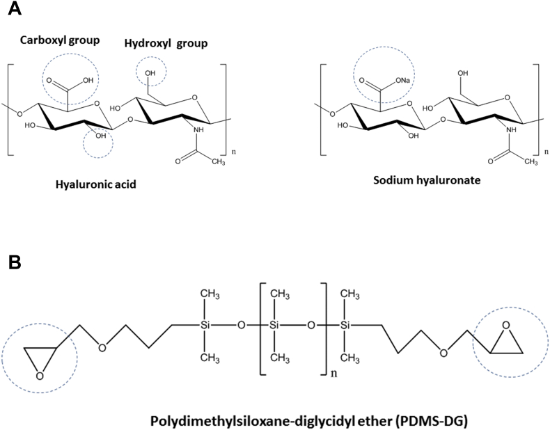
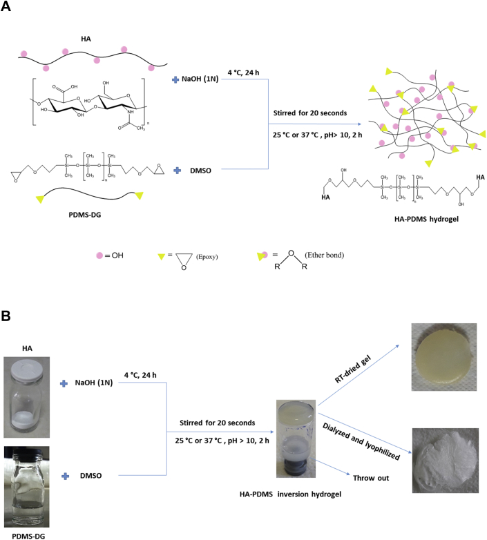
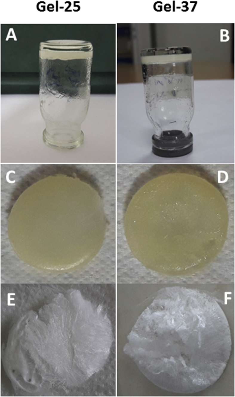
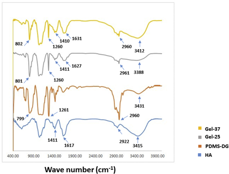
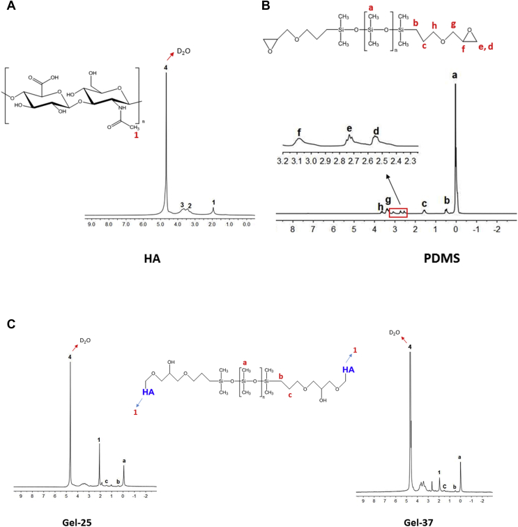
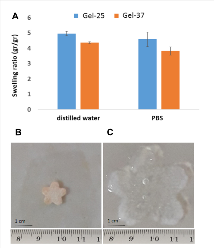
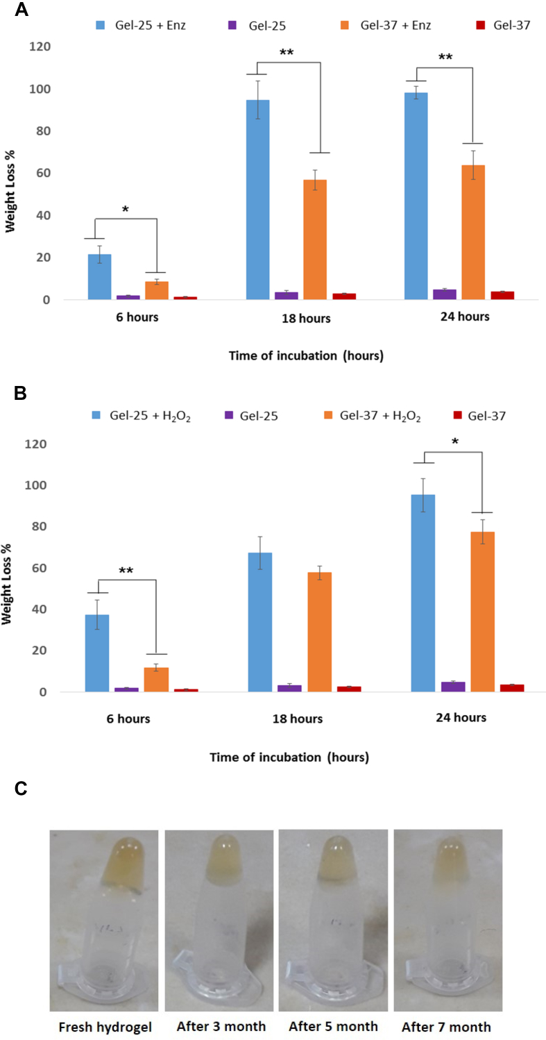
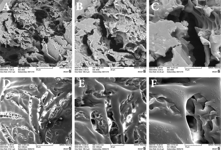
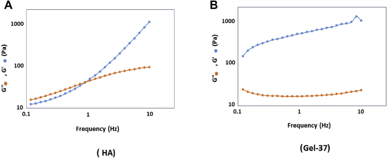
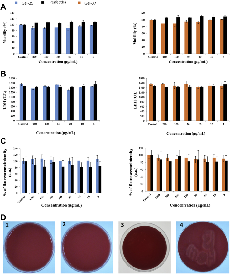
Hyaluronic acid (HA), as a safe biomaterial with minimal immunogenicity, is being employed in a broad range of medical applications. Since unmodified HA has a high potential for biodegradation in the physiological condition, herein, an HA-based cross-linked hydrogel was formulated using polydimethylsiloxane-diglycidyl ether terminated (PDMS-DG) via epoxide-OH reaction. The formation of HA-PDMS hydrogel was confirmed using FTIR, NMR, and FESEM. Temperature demonstrated a critical role in the physicochemical properties of the final products. Gel-37, which formed at 37 °C, had a higher modification degree (MD) and more stability against hyaluronidase and oxidative stress than the hydrogel formulated at 25 °C (Gel-25). In addition, the swelling ratio, roughness, and porous network topology of Gel-25 and Gel-37 were different. The rheology measurement indicated that HA-PDMS hydrogel had a stable viscoelastic character. The hydrogel was also biocompatible, non-cytotoxic, and considerably stable during 7-months storage. Overall, various determined parameters confirmed that HA-PDMS hydrogel is worth using in different medical applications. Keywords: Hyaluronic acid; Polydimethylsiloxane-diglycidyl ether terminated; Hydrogels; Long-term stability; Viscoelastic behavior; Biocompatibility.

Keywords: Biocompatibility; Hyaluronic acid; Hydrogels; Long-term stability; Materials science; Polydimethylsiloxane-diglycidyl ether terminated; Viscoelastic behavior.


Figures

Figure 1
Figure 1
Chemical structure of HA and Sodium hyaluronate (A) and PDMS-DG (B). Two functional groups, hydroxyl and carboxyl, are shown by blue circles. Sodium hyaluronate is dominant form of HA at physiological conditions. Polydimethylsiloxane has two methyl groups attached to its silicon structure. PDMS-DG has two epoxy groups in its ends (blue circles). The chemical structures present here have been drawn with ChemBioDraw Uitra 12.0.
Figure 2
Figure 2
HA-PDMS hydrogel synthesis at different temperatures. (A) Schematic diagram and (B) real materials representation. HA was incubated at basic pH, and interaction of the epoxide groups with the HA hydroxyl groups leads to ether bond formation. This cross-linking reaction created HA-PDMS 3D hydrogel network.
Figure 3
Figure 3
The hydrogels producing at 25 °C (A, C, E) and 37 °C (B, D, F). Inversion property of the gels was tested by inverting the vessels after removing the non-reacted materials (A, B). The dried gels at room temperature, or the dialyzed/lyophilized gels are represented in (C, D) and (E, F), respectively.
Figure 4
Figure 4
FTIR spectra of HA, PDMS-DG, Gel-25, and Gel-37. The characteristic peaks of HA and PDMS-DG were indicated by blue arrows, which also presented in the constructed hydrogels.
Figure 5
Figure 5
1H NMR spectra of HA, PDMS-DG, Gel-25, and Gel-37. (A) The HA spectrum in D2O with specific peaks for the N-acetyl group (1) and sugar rings (2, 3). (B) The PDMS-DG spectrum in chloroform with specific peaks for the methyl (a), epoxy (d, e, f), and CH2 (b, c, g, h) groups. (C) The Gel-25 and the Gel-37 spectra in D2O with the specific peaks of HA and PDMS-DG. The red number or letters in the chemical structures represented the chemical groups detected by NMR.
Figure 6
Figure 6
Assessment of the swelling properties of the hydrogels. (A) The swelling ratio of Gel-25 and Gel-37 in distilled water and PBS at 37 °C. (B) A designed pluck of Gel-37 after dried in room temperature and (C) after re-hydration.
Figure 7
Figure 7
Assessing the stability of the hydrogels against degradation. (A) Enzymatic degradation of Gel-25 and Gel-37 using 7 U/mL hyaluronidase at 37 °C. (B) Chemical degradation of the Gel-25 and Gel-37 using 2.5 % (v/v) H2O2 at 37 °C. The control samples were incubated in PBS without any enzyme or H2O2 (N = 3, mean ± SD, ∗P < 0.05, ∗∗P < 0.01). (C) Follow up the inversion property of Gel-37 after seven months staying at 25 °C.
Figure 8
Figure 8
FESEM micrographs of the hydrogels. A, B, and C are for Gel-25 and D, E, and F are for Gel-37. Imaging was performed after coating the lyophilized hydrogels with gold. The detail of magnification and scale bars are demonstrated under each image.
Figure 9
Figure 9
Distribution of the elements in the hydrogel, EDS curve of Gel-37. The inset indicated the percentage of each element, including O, C, N, Na, and Si.
Figure 10
Figure 10
Distribution of the elements in the hydrogel, Elemental mapping (EM) profile of Gel-37. All of the Na-HA and PDMS-DG chemical elements (C, O, N, Na, and Si) except H are visible in the hydrogel.
Figure 11
Figure 11
Rheological evaluating of the hydrogel. Logarithmic variations of the storage module, G′, and the loss module, G″, of (A) pure HA and (B) Gel-37 based on the loading frequency.
Figure 12
Figure 12
Cytotoxicity and biocompatibility measurements of Gel-25 and Gel-37. The viability of L929 fibroblastic cell line treated with different concentrations of the hydrogels for 24 h, were analyzed using (A) MTT and (B) LDH assays. (C) Evaluation of the ROS generation in the cells by measuring the DCF fluorescence intensity in the presence of different concentrations of Gel-25, Gel-37, or Perfectha. (D) Biocompatibility assay using the hemolysis test, Gel-25 (1), Gel-37 (2) or Perfectha (3), and Bacillus Cereus as a positive control (4).

Similar articles

Cited by

References

    1. Al-Sibani M., Al-Harrasi A., Neubert R.H. Study of the effect of mixing approach on cross-linking efficiency of hyaluronic acid-based hydrogel cross-linked with 1, 4-butanediol diglycidyl ether. Eur. J. Pharmaceut. Sci. 2016;91:131–137. - PubMed
    1. Fakhari A., Berkland C. Applications and emerging trends of hyaluronic acid in tissue engineering, as a dermal filler and in osteoarthritis treatment. Acta Biomater. 2013;9:7081–7092. - PMC - PubMed
    1. Schanté C.E., Zuber G., Herlin C., Vandamme T.F. Chemical modifications of hyaluronic acid for the synthesis of derivatives for a broad range of biomedical applications. Carbohydr. Polym. 2011;85:469–489.
    1. Kablik J., Monheit G.D., Yu L., Chang G., Gershkovich J. Comparative physical properties of hyaluronic acid dermal fillers. Dermatol. Surg. 2009;35:302–312. - PubMed
    1. Robert L., Robert A.-M., Renard G. Biological effects of hyaluronan in connective tissues, eye, skin, venous wall. Role in aging. Pathol. Biol. 2010;58:187–198. - PubMed

LinkOut - more resources