Review
. 2020 Apr 5:5:25.
doi: 10.21037/tgh.2019.11.15. eCollection 2020.

Inherited iron overload disorders

Affiliations
Review

Inherited iron overload disorders

Alberto Piperno et al. Transl Gastroenterol Hepatol. .

Abstract

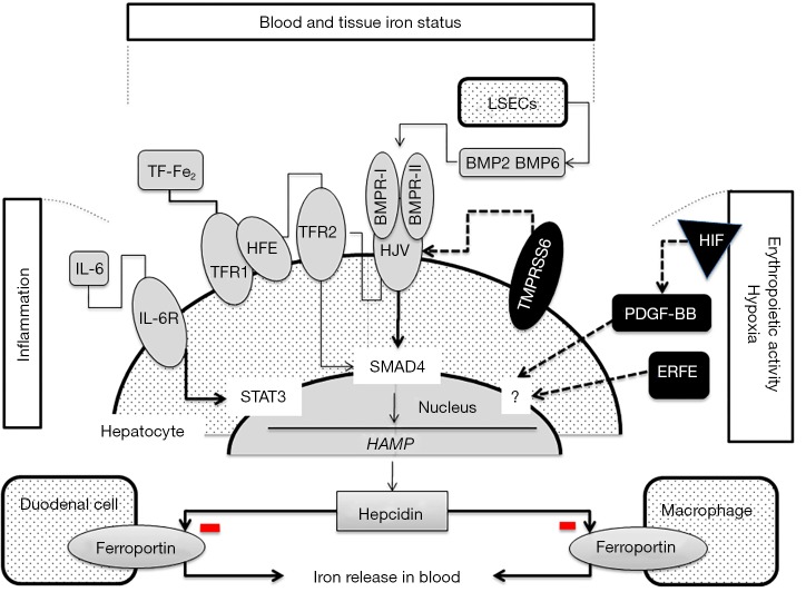
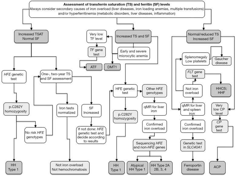
Hereditary iron overload includes several disorders characterized by iron accumulation in tissues, organs, or even single cells or subcellular compartments. They are determined by mutations in genes directly involved in hepcidin regulation, cellular iron uptake, management and export, iron transport and storage. Systemic forms are characterized by increased serum ferritin with or without high transferrin saturation, and with or without functional iron deficient anemia. Hemochromatosis includes five different genetic forms all characterized by high transferrin saturation and serum ferritin, but with different penetrance and expression. Mutations in HFE, HFE2, HAMP and TFR2 lead to inadequate or severely reduced hepcidin synthesis that, in turn, induces increased intestinal iron absorption and macrophage iron release leading to tissue iron overload. The severity of hepcidin down-regulation defines the severity of iron overload and clinical complications. Hemochromatosis type 4 is caused by dominant gain-of-function mutations of ferroportin preventing hepcidin-ferroportin binding and leading to hepcidin resistance. Ferroportin disease is due to loss-of-function mutation of SLC40A1 that impairs the iron export efficiency of ferroportin, causes iron retention in reticuloendothelial cell and hyperferritinemia with normal transferrin saturation. Aceruloplasminemia is caused by defective iron release from storage and lead to mild microcytic anemia, low serum iron, and iron retention in several organs including the brain, causing severe neurological manifestations. Atransferrinemia and DMT1 deficiency are characterized by iron deficient erythropoiesis, severe microcytic anemia with high transferrin saturation and parenchymal iron overload due to secondary hepcidin suppression. Diagnosis of the different forms of hereditary iron overload disorders involves a sequential strategy that combines clinical, imaging, biochemical, and genetic data. Management of iron overload relies on two main therapies: blood removal and iron chelators. Specific therapeutic options are indicated in patients with atransferrinemia, DMT1 deficiency and aceruloplasminemia.

Keywords: Iron overload; ferritin; transferrin saturation.


Conflict of interest statement

Conflicts of Interest: The authors have no conflicts of interest to declare.

Figures

Figure 1
Figure 1
Positive and negative regulators of hepcidin synthesis. Hepcidin regulators are activated by different stimuli: positive (iron status, inflammation), and negative (erythropoietic activity, and hypoxia). HFE is a component of an iron-sensing complex that involves interactions with diferric transferrin (Tf-Fe2), transferrin receptors (TFR-1 and TFR-2) at the plasma membrane of hepatocytes. High concentrations of Tf-Fe2 displace HFE from TFR1, which then forms a complex with TFR2 and HJV to promote bone morphogenetic protein (BMP)/SMAD signaling to hepcidin (28,29). HJV is a GPI-linked protein that activates hepcidin as a co-receptor for BMP cytokines. Only BMP2 and BMP6 have been so far demonstrated to activate hepcidin in vivo (9,21,30,31). In the liver, hepcidin is expressed only in hepatocytes while BMP2 and BMP6 are expressed almost exclusively in liver sinusoidal endothelial cells (LSECs) suggesting a paracrine function of these ligands (32). How LSECs sense changes in body iron levels and upregulate BMP2 and BMP6 is still to be defined. Two types of BMP receptors, type I (BMPRI) and type II (BMPRII) are involved in this pathway, and both types, as well as their ligands, act as dimers. The regulatory SMADs (R-SMADs), SMAD1, SMAD5, and SMAD8 are the mediators of BMP signalling, as they are phosphorylated by activated BMPRIs. The common SMAD4 translocates to the nucleus in complex with the R-SMADs to induce the expression of genes regulated by BMP-responsive elements, as hepcidin. BMP/SMAD signaling to hepcidin is suppressed by matriptase 2, a serine protease, codified by TMPRSS6 that cleaves and generates a soluble form of HJV. Erythroferrone (ERFE), a TNFα-like protein released by mature erythroblasts in condition of enhanced erythropoiesis, is a major candidate of erythropoiesis-induced hepcidin suppression (33). Platelet-derived growth factor-BB (PDGF-BB) is the candidate of hypoxia-induced hepcidin inhibition by the hypoxia-inducible factor (HIF), likely produced by a non-erythroid Ter119neg population under hypoxia stimuli (34,35). Infection and inflammation markedly increase hepcidin synthesis through the interaction of interleukin 6 (IL6) with its receptor (IL6R) and STAT3 pathway (36).
Figure 2
Figure 2
Diagnostic flow-chart for hereditary iron overload disorders. A careful evaluation for secondary iron overload or hyperferritinemia should always be done (see text). High TSAT and SF in pediatric patients with severe microcytic anemia might imply several iron loading anemia including congenital atransferrinemia and DMT1 deficiency. A TSAT cut-off value of 45% is commonly chosen in phenotypic screening for hemochromatosis. Upper normal values for serum ferritin (SF) differ according to age and gender and in adults vary from 300 to 400 µg/L in men and from 160 to 250 µg/L in pre-menopausal and post-menopausal women, respectively (161,162). Compound heterozygosity for p.Cys282Tyr/p.His63Asp and, even more, homozygosity for p.His63Asp have very low penetrance and expression, and patients with these genotypes should be evaluated with caution, carefully considering the presence of other causes of iron overload and hyperferritinemia. p.Cys282Tyr heterozygotes with hemochromatosis phenotype can be evaluated for rare mutations in HFE or in other hemochromatosis genes. Patients with confirmed high TSAT with still normal SF may undergo to genetic ascertainment or 1–2-year follow-up. Quantitative iron assessment by magnetic resonance (qMR) or, more rarely, liver biopsy, is mandatory in suspected non-HFE hemochromatosis. In patients with hyperferritinemia with normal/low ceruloplasmin (Cp) should be measured and family study is helpful in the diagnostic approach of autosomal dominant form of hyperferritinemia as ferroportin disease, hereditary-hyperferritinemia-cataract syndrome (HHCS) and benign hyperferritinemia (HHF). Patients with ferroportin disease might show a strong signal in spleen qMR related to the prevalent reticuloendothelial iron accumulation. Hyperferritinemia with even mild splenomegaly and thrombocytopenia can give rise to Gaucher’s disease suspicion.

References

    1. Cook A, Giunti P. Friedreich's ataxia: clinical features, pathogenesis and management. Br Med Bull 2017;124:19-30. 10.1093/bmb/ldx034 - DOI - PMC - PubMed
    1. Iolascon A, De Falco L, Beaumont C. Molecular basis of inherited microcytic anemia due to defects in iron acquisition or heme synthesis. Haematologica 2009;94:395-408. 10.3324/haematol.13619 - DOI - PMC - PubMed
    1. Levi S, Rovida E. Neuroferritinopathy: From ferritin structure modification to pathogenetic mechanism. Neurobiol Dis 2015;81:134-43. 10.1016/j.nbd.2015.02.007 - DOI - PMC - PubMed
    1. Furuyama K, Kaneko K. Iron metabolism in erythroid cells and patients with congenital sideroblastic anemia. Int J Hematol 2018;107:44-54. 10.1007/s12185-017-2368-0 - DOI - PubMed
    1. Girelli D, Bozzini C, Zecchina G, et al. Clinical, biochemical and molecular findings in a series of families with hereditary hyperferritinaemia-cataract syndrome. Br J Haematol 2001;115:334-40. 10.1046/j.1365-2141.2001.03116.x - DOI - PubMed