. 2020 Mar 3;5(12):6763-6772.
doi: 10.1021/acsomega.0c00098. eCollection 2020 Mar 31.

Cane Molasses Graphene Quantum Dots Passivated by PEG Functionalization for Detection of Metal Ions

Affiliations

Cane Molasses Graphene Quantum Dots Passivated by PEG Functionalization for Detection of Metal Ions

Ying Lou et al. ACS Omega. .

Abstract

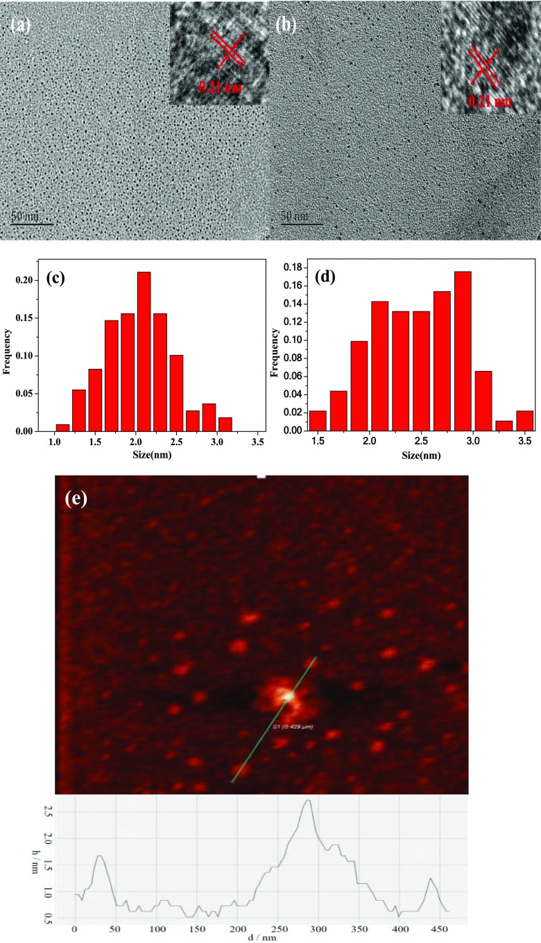
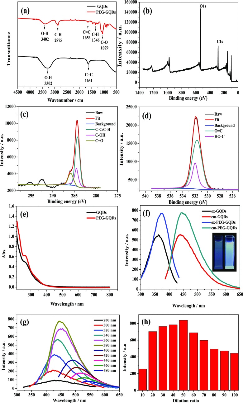
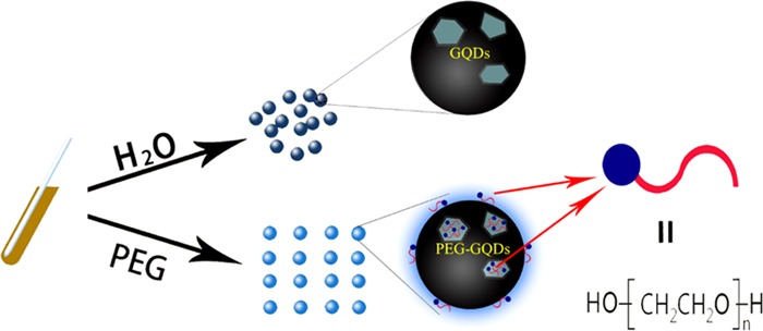
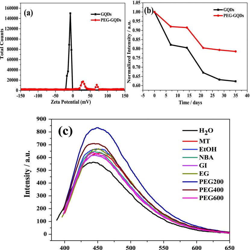
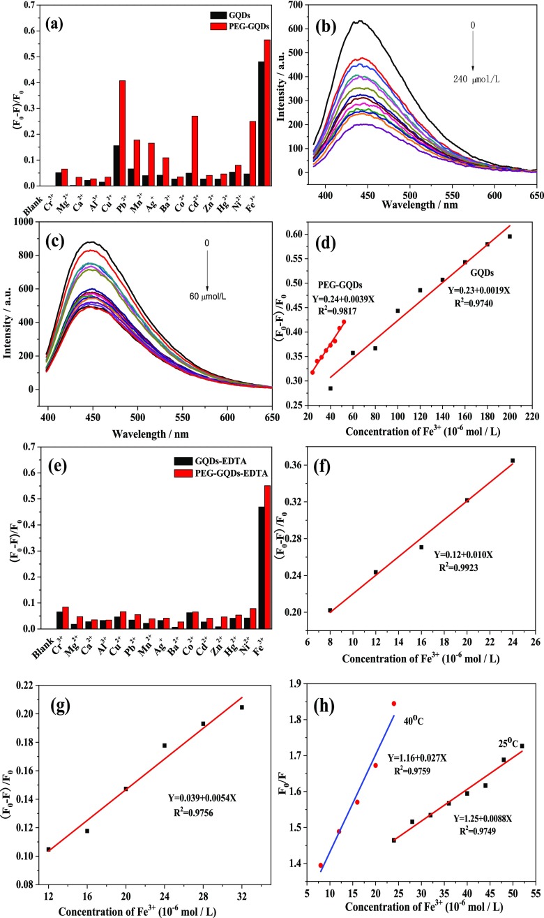
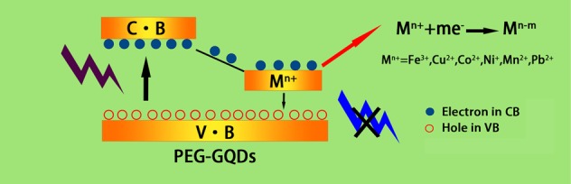
Poly(ethylene glycol) passivated graphene quantum dots (PEG-GQDs) were synthesized based on a green and effective strategy of the hydrothermal treatment of cane molasses. The prepared PEG-GQDs, with an average size of 2.5 nm, exhibit a brighter blue fluorescence and a higher quantum yield (QY) (up to approximately 21.32%) than the QY of GQDs without surface passivation (QY = 10.44%). The PEG-GQDs can be used to detect and quantify paramagnetic transition-metal ions including Fe3+, Cu2+, Co2+, Ni2+, Pb2+, and Mn2+. In the case of ethylenediaminetetraacetic acid (EDTA) solution as a masking agent, Fe3+ ions can be well selectively determined in a transition-metal ion mixture, following the lowest limit of detection (LOD) of 5.77 μM. The quenching mechanism of Fe3+ on PEG-GQDs belongs to dynamic quenching. Furthermore, Fe3+ in human serum can be successfully detected by the PEG-GQDs, indicating that the green prepared PEG-GQDs can be applied as a promising candidate for the selective detection of Fe3+ in clinics.


Conflict of interest statement

The authors declare no competing financial interest.

Figures

Figure 1
Figure 1
TEM and HRTEM images of (a) GQDs and (b) PEG-GQDs; size distribution of (c) GQDs and (d) PEG-GQDs; and (e) atomic force microscopy (AFM) image of GQDs.
Figure 2
Figure 2
(a) FT-IR spectra of GQDs and PEG-GQDs. (b) X-ray photoelectron spectroscopy (XPS) survey scan spectrum of PEG-GQDs. High-resolution XP spectra of (c) carbon and (d) oxygen of PEG-GQDs. (e) UV–vis absorption spectra of GQDs and PEG-GQDs. (f) Fluorescence emission spectra of GQDs and PEG-GQDs and the inset of the fluorescence photographs of GQDs (left) and PEG-GQDs (right). (g) Fluorescence emission spectra of PEG-GQDs recorded for progressively longer excitation wavelengths from 280 to 480 nm. (h) Diameter histogram of the effect of adding an amount of PEG on the fluorescence emission spectra of PEG-GQDs.
Figure 3
Figure 3
Schematic illustration of the function of PEG for GQDs.
Figure 4
Figure 4
(a) ζ-potentials of GQDs and PEG-GQDs. (b) Stability of GQDs and PEG-GQDs for 35 days. (c) Fluorescence emission spectra of passivated GQDs under different modifiers.
Figure 5
Figure 5
(a) Fluorescence responses of GQDs and PEG-GQDs to the different metal ions in the absence of ethylenediaminetetraacetic acid (EDTA). (b) Fluorescence emission spectra of GQDs quenched by different concentrations of Fe3+ (from 0 to 240 μM) in a homogeneous solution. (c) Fluorescence emission spectra of PEG-GQDs quenched by different concentrations of Fe3+ (from 0 to 60 μM) in a homogeneous solution. (d) Calibration curves of the degree of fluorescence quenching [(F0F)/F0] of GQDs and PEG-GQDs versus Fe3+ ions concentration. (e) Fluorescence responses of GQDs and PEG-GQDs to different metal ions in the presence of EDTA. Calibration curves of the degree of fluorescence quenching [(F0F)/F0] of (f) PEG-GQDs and (g) GQDs versus Fe3+ ions concentration in the presence of EDTA, respectively. (h) Stern–Volmer curve for fluorescence quenching of PEG-GQDs by Fe3+ at different temperatures.
Figure 6
Figure 6
Quenching mechanism of metal ions of PEG-GQDs.

Similar articles

Cited by

References

    1. Sharma V.; Tiwari P.; Mobin S. M. Sustainable carbon-dots: recent advances in green carbon dots for sensing and bioimaging. J. Mater. Chem. B 2017, 5, 8904–8924. 10.1039/C7TB02484C. - DOI - PubMed
    1. Ju B.; Wang Y.; Zhang Y. M.; Zhang T.; Lu Z. H.; Li M. J.; Zhang S. X. A. Photostable and Low-Toxic Yellow-Green Carbon Dots for Highly Selective Detection of Explosive 2,4,6-Trinitrophenol Based on the Dual Electron Transfer Mechanism. ACS Appl. Mater. Interfaces 2018, 10, 13040–13047. 10.1021/acsami.8b02330. - DOI - PubMed
    1. Hu X. H.; An X. Q.; Li L. L. Easy synthesis of highly fluorescent carbon dots from albumin and their photoluminescent mechanism and biological imaging applications. Mater. Sci. Eng. C 2016, 58, 730–736. 10.1016/j.msec.2015.09.066. - DOI - PubMed
    1. Pan J. Q.; Sheng Y. Z.; Zhang J. X.; Wei J. M.; Huang P.; Zhang X.; Feng B. X. Preparation of carbon quantum dots/TiO2 nanotubes composites and their visible light catalytic applications. J. Mater. Chem. A 2014, 2, 18082–18086. 10.1039/C4TA03528C. - DOI
    1. Wang L. P.; Wu X. Q.; Guo S. J.; Han M. M.; Zhou Y. J.; Sun Y.; Huang H.; Liu Y.; Kang Z. H. Mesoporous nitrogen, sulfur co-doped carbon dots/CoS hybrid as an efficient electrocatalyst for hydrogen evolution. J. Mater. Chem. A 2017, 5, 2717–2723. 10.1039/C6TA09580A. - DOI